http://www.mindfully.org/Technology/Microwave-Effects-Goldsmith1dec97.htm
Department of Epidemiology and Health Services Evaluation, Ben-Gurion University of the Negev, Beer Sheva, Israel
Medline: Goldsmith JR. Epidemiologic evidence relevant to radar (microwave) effects. Environ Health Perspect. 1997 Dec;105 Suppl 6:1579-87. Review. PMID: 9467086 [PubMed - indexed for MEDLINE]
Key words: leukemia epidemiology, brain cancer epidemiology, nonionizing radiation epidemiology, cellular telephones and health, TV and radio broadcast towers, military electronic equipment effects
This paper is based on a presentation at the International Conference on Radiation and Health held 3-7 November 1996 in Beer Sheva, Israel. Abstracts of these papers were previously published in Public Health Reviews 24(3-4):205-431 (1996). Manuscript received at EHP 11 March 1997; accepted 2 June 1997.Address correspondence to Dr. J.R. Goldsmith, Department of Epidemiology and Health Services Evaluation, Faculty of Health Sciences, Ben-Gurion University of the Negev, P.O. Box 653, Beer Sheva, Israel 84120. Telephone: 972 7 6400876. Fax: 972 7 6277342. E-mail: gjohn@bgumail.bgu.ac.il
Abbreviations used: AE, aviation electrician's mate(s); ALL acute lymphatic leukemia; ANLL, acute nonlymphatic leukemia; AT, aviation electronics technician(s); ELF, extremely low frequency radiation (50-60 cycles); FM, frequency modulation; FSHSS, Foreign Service Health Status Study (Lilienfeld Report); FT, fire control technician(s); O/E, observed to expected; OR, odds ratio(s); RD, radarmen; RF, radiofrequency or microwave; RM, radiomen; RR, relative risk; SIR, standardized incidence ratios; TV, television; U.S. EPA, U.S. Environmental Protection Agency; UV, ultraviolet.
ELF studies have included extensive evaluations of occupational and residential exposures, but there has been considerable difficulty in establishing dose-response relationships or mechanisms (7).
Evaluation of RF exposures was conducted primarily by military- and security-oriented government agencies, and earlier studies recently have been reevaluated. Because of the rapid development and use of cellular telephone systems, which involve widespread public exposures, reevaluation of exposure risk becomes urgent. The radiation emanates mainly from handheld devices and from the many broadcast facilities needed to maintain such systems. It is generally agreed that the physical attributes of UV, ELF, and RF exposures are sufficiently different so that separate evaluations of the possible risks of each are justified. Nevertheless, some potential mechanisms of biologic reaction and dose-response relationships among different types of subjects and exposures are common to all three exposures. I discuss the types of epidemiologic evidence with possible relevance to evaluation of RF exposures.
Neoplastic responses, if they do occur, may have a long latency period. If one waits for cancer to occur, exposed populations have increased risk for the duration of the latency period. Even if cancer risks cannot yet be unequivocally demonstrated, some measure of protection should be taken as early as possible because it may take some time to determine the definitive relationship between RF and cancer.
A second reason for urgency with respect to taking protective measures is that, because of rapid increases in the numbers of persons exposed to increased RF exposures in connection with cellular telephone use, some biologic basis is needed as a guide for prudent protective behavior. It is possible that a system of biologic indicators can be found that would allow identification of increased cancer risk. This possibility seems worth exploring.
Originally, heating of tissues by RF was considered the basic mechanism through which radiation affected exposed individuals. Therefore, existing protective principles and practices are built around avoidance of the thermal effects of such exposures. There has been increasing concern that this approach may not be adequate and with this in mind, the International Conference on Non-Thermal Effects of Microwave Radiation was convened in November 1996. The proceedings of this conference are being prepared for publication.
With regard to epidemiologic evidence of radiation, a report published in January 1995 (8) focused primarily on military, industrial, and broadcast exposures. This study was supplemented by the review of Rothman et al. (9) in May 1996, Grayson's report on brain cancer in U.S. Air Force personnel (10), and by three published community studies of cancers possibly associated with proximity to broadcast facilities (11-13).
This review is intended only to update previous assessments of cancer risk from RF radiation. Like the earlier report (8), it is not intended to be a comprehensive review or a balanced report of all possibly relevant findings. Also, there is no attempt to critically evaluate these findings.
Finally, Swedish government agency assessments of ELF effects led to prudent avoidance policies (14,15), which may be applicable to presently available knowledge about RF. This appears to be an attractive alternative to waiting until the data are convincing enough to achieve agreement on new and enforceable standards.
Evidence up to 1994 as reviewed in Goldsmith (8) indicates the likelihood of the following effects from exposure to RF radiation in certain populations: reproductive effects such as increased spontaneous abortion, changes in blood counts, increased somatic mutation, and increased incidence of childhood and other cancers. Other findings have suggested effects such as cataract, nonspecific disabilities, and symptoms in sensitive persons (headache, ocular problems, fatigue, dizziness, memory impairment, and sleep difficulties).
Reproductive Outcomes
Study of Physiotherapists. Ouellet-Hellstrom and Stewart (16) reported on a study of female physiotherapists who used either RF or short-wave apparatus and were queried about the outcome of pregnancies. The frequency generated by short-wave equipment was 27.12 MHz and by microwave equipment was 915 MHz and 2450 MHz.
The survey was conducted among female members of the American Physiotherapy Association in the United States. Of 11,598 respondents who reported having at least one pregnancy, 6684 (57.6%) reported using short-wave or microwave diathermy. These 6684 women reported 14,989 pregnancies, of which 1791 ended in miscarriages, called case pregnancies. Of the remaining pregnancies with sufficient data, 12,949 were classified as control pregnancies. Exposure was defined as occurring if the woman had been using one of the physiotherapy modes during the first trimester of pregnancy and during the preceding 6 months. Cases were matched to controls by mother's age at time of conception and by the number of years elapsed between the pregnancy and the date of filling out the questionnaire. A number of confounders were included, among which was prior fetal loss.
Of the case and control mothers, 11.9 and 9.5%, respectively, were using microwaves during the pregnancy; the odds ratio (OR) for spontaneous abortion increased as the number of exposures increased from 5 or less to 20 or more per month. The trend was significant whether or not prior fetal loss had occurred. For women exposed to short-wave radiation, 22.3% lost their baby prior to the 7th week of pregnancy, whereas the figure for unexposed women was 24.4%. Of the microwave-exposed women, 47.7% had miscarriages prior to the 7th week of pregnancy compared to 14.5% of nonexposed women.
Measured values of stray emissions near waist level ranged from 0.04 to 16.58 mW/cm2 for electric fields with short-wave units, and these units produced magnetic fields of 0.09 to 8.32 mW/cm2. For microwave diathermy the electric field leakage was from 0.08 to 1.20 mW/cm2. Leakage measured 15 cm from the source was as high as 15 mW/cm2. Duration of the therapist exposures was usually only a few minutes per treatment.
Moscow Staff Study. The exposures of U.S. embassy personnel in Moscow are described in Goldsmith (8), based on Lilienfeld at al. (17) (Table 1). Studies were done among Moscow embassy employees, staff dependents, and other personnel and compared with similar groups in other Eastern European embassies.
Table 1. Complications of pregnancy, childbirth, and puerperium (ICD-8, Codes 630—678) among women employees in the Foreign Service Health Status Study (17).
		Evera,b				After indexa,c
        Moscow	  Comparison 		Moscow     Comparison 		p
	19 (6%)   19 (3%)    		11 (3.5%)  9 (1.3%)   		0.04
SMBR    1.7       0.67
Abbreviations: ICD. International Classification of Diseases, 8th Revision: SMBR, standardized morbidity ratio. a Refers to the initial tours of duty during which exposures occurred. b Whether the condition occurred at any time; 'Whether the condition occurred after the initial tour of duty.
The study known as the Foreign Service Health Status Study (FSHSS) or Lilienfeld Report (17) was designed to compare the experience of employees in the Moscow embassy with those of similar employees in other Eastern European embassies on the assumption that the latter were not exposed to RF radiation. There was some evidence that these employees were exposed as well, but the contract officer dismissed the possibility as being based on hearsay. In a meeting with the State Department Contract Officer Dr. Pollack about the submitted draft of the Lilienfeld Report, G. Jacobsonnoted that the reference to a potential infertility effect in the study might be inappropriate because the experimental work was done at very high doses and there are no controlled human studies (18). According to the minutes of the meeting, "this clause will be modified to reflect the very speculative nature of the reports, but the FSHSS data will be presented as is" (17).
The final report makes no reference to any possible impact on infertility, but it does present some data (Table 1) that show more frequent complications among Moscow workers compared to those from other embassies.
Thus, we are left with higher rates of complications of pregnancy at the Moscow embassy for a problem that originally was thought to affect fertility. It seems most likely related to or actually to be spontaneous abortion.
Systematic Alterations in Red or White Blood Cell Counts
When radar was first identified as a health risk, Daily (19) reported a statistically significant increase in immature red blood cells among workers exposed to radar. These studies were summarized by Follis et al. (20). Early studies at Lockheed Aircraft (Burbank, CA) by Barron et al. (21) were later dismissed on the grounds "that there was variation in the interpretations by a laboratory technician" (22). Bach found that rats exposed at 13 mW/cm2 had changes in blood cell counts (23).
Table 2. Results of tests for chromosomal changes in metaphase spreads of lymphocytes cultured in vitro among selected Moscow embassy employees.
Mutagenic levela 	Designator	Subjects, no
5			Extreme		0
4			Severe		6
3.5			Intermediate	5
3			Moderate	7
2.5			Intermediate	5 -
2			Questionable	5
1			Normal		6
Growth failure				2
a Grading of mutagenic processes and clinical interpretations of these findings were provided by Dr. G. Jacobson (George Washington University Medical School, Washington, DC), who wrote: "Patients who repeat at level 3 or higher should not reproduce until 6 months after somatic levels have returned to 2 or 1. Patients at level 4 should be withdrawn from mutagenic exposure and monitored each month until less than 3 is obtained on two consecutive samples' (18). Dr. Jacobson also wrote, feel impelled, as in past reports, to emphasize the necessity to study serial samples on the same individual and when possible to study the subject prior to exposure" 1181. Apparently, no such follow-up or serial studies were done.
Goldoni (24,25) compared the hematological findings in 25 male air traffic control technicians exposed to radar with those for 10 electronic technicians whose work was distant from a microwave source. The radar was in the range of 1250 to 1350 MHz, with a strength varying from 10 to 20 µW/cm2 . Radar-exposed workers had significantly lower levels of leukocytes and red cells than workers distant from the microwave source. In a follow-up study of 49 radar-exposed technicians, thrombocyte and leukocyte counts decreased significantly but stayed within normal limits (25).
A hematologic study of Moscow foreign service workers was submitted to the U.S. government on 7 October 1976 by Tonascia and Tonascia (26). They found, on comparing the data for Moscow-based employees with that from foreign service exams conducted in the United States, that [mindfully.org note: end of sentence missing in online version]
The differences between the two groups with respect to every parameter except monocytes (% and counts) are highly statistically significant (p<0.001) after appropriate transformation. Specifically the Moscow group had a higher mean hematocrit, the Moscow group had a lower neutrophil percentage, but higher percentages for the other three cell types (lymphocytes, eosinophils, and monocytes). The white cell counts are strikingly higher in the Moscow group.
Several statistically significant changes occurred over time in the Moscow group; specifically, mean hematocrit increased and a 3-fold increase in monocyte count occurred. Neutrophil percentages fell and then rose; the reverse pattern was observed for the lymphocytes (26).
Vukelic et al. (27) studied the effects of RF radiation on 72 physiotherapists and physiatrists in Croatia. They found a significantly positive correlation between length of service and white cell count, and an association of years of exposure with low red cell count.
Tornqvist et al. (28) studied 706 power station workers at 3-year intervals and found that the white blood cell counts were decreased slightly because of exposures to magnetic fields.
Evidence of Mutational Activity in Human Incubated White Blood Cells
The initial examination of Moscow embassy workers, conducted when it became known they were being irradiated by Soviet transmitters, was done to study the possible effects of radiation on chromosomes in blood samples (26). Beginning in February 1966, 3 to 4 years after the microwave irradiation was first detected, samples were taken for chromosomal analysis. Twenty spreads were scored per sample; results are shown in Table 2 (18).
Overexposed Air Traffic Controllers. Garaj-Vrohac et al. (29) examined six men accidentally exposed while repairing microwave devices used for air traffic control in Zagreb. These subjects usually worked alternate days in a microwave field of 1250 to 1350 MHz with power density of 10 µg/W/cm2 to 20 mW/cm2. The accidental exposure was greater than these figures but by how much is not known. The results of chromosome aberration analysis during 1984 to 1990 showed no increase in chromosomal abnormalities compared to the control. Table 3 shows results for the accidentally exposed subjects.
Table- 3. Type and percentage of chromosomal aberrations after accidental exposure to high-power density pulse RF radiation.
Subject-       	   Chromatid  Chromosome                                Total
  no      Date     breaks     breaks  	Acentrics Dicentrics Rings   aberrations, %
  1     18/11/1990  2          1      	  2          1         —         3.0
         5/12/1990  —          1      	  1          1         —         1.5
  2      6/12/1990  —          4      	  8          4         1         8.5
        25/02/1991  4          1      	  3          1         —         4.5
  3      6/12/1990             3      	 10          3         —         8.5
        26/02/1991  4          3      	  3          2         —         6.0
  4     20/12/1990  —          1      	  2                    1         2.0
        16/01/1991  —          3          5          1         1         5.0
  5     11/12/1990  —          6      	 48          9         3        33.0
        14/02/1991  1          4      	 31          6         2        22.0
        13/03/1991  4          7      	 18          6                  18.0
        17/04/1991  6          6      	  6          a         a         9.5
        22/05/1991  3          4      	  6          2         —         7.5
         2/06/1991  1          —      	  5          1         —         3.5
  6     20/12/1990  —          4     	  2          1         —         3.5
        30/01/1991  —          2       	  1          —         1         2.0

  a  Chromatid interchange 1.
Two things are clear from this experience: Microwave irradiation can produce genotoxic effects, and recovery can occur with a half-time of about 15 weeks when about one-third of the spreads show aberrations. Both chromosomal and chromatid reactions occur.
It is conventional wisdom to assume that nonionizing radiation cannot produce such changes, but there is evidence that this view is incorrect. For example, cattle in the field exposed in vivo near a large military RF emitter in Skrunda, Latvia (30), showed more positive micronuclei test results than unexposed cattle. Bovine lymphocytes in vitro respond to microwave exposure using the same test (31). Genotoxic changes are found in Chinese hamster cells in vitro (thymidine incorporation and chromosomal and chromatid changes) (32) and in human lymphocytes in vitro (33) using micronuclei tests.
A series of studies from Croatia and Italy have also demonstrated that radar exposures are mutagenic both in vivo and in vitro (29,32-35).
In a paper about the effect of RF radiation on the cell genome (32), the investigators used cultured Chinese hamster cells exposed to 7.7 GHz at power densities of 30 mW/cm2 for 15, 30, and 60 min. Using tritiated thymidine and autoradiography, the incorporation of thymidine into DNA after a 4-hr incubation decreased in a stepwise manner according to the length of exposure and almost completely recovered in 24 hr. In addition, chromosomal aberrations increased stepwise according to the duration of exposure. The background percent abnormal metaphase was 1.7%; with a 15-min exposure it increased to 4.8%, with 30 min, 6.3%, and with 60 min, 8.9%. Garaj-Vhrovac et al. (33) report on the relationship between colony-forming ability, chromosome aberrations, and the incidence of micronuclei in V79 Chinese hamster cells exposed to RF radiation. These authors were able to demonstrate damage to cell genomes and changes in chromosome structure based on observations of structural chromosomal aberrations and micronuclei tests. The exposures used were 7.7 GHz and 30 mW for 15, 30, and 60 min. The structural changes replicated the changes observed in their initial paper. The micronuclei/1000 cells were background 0.016, and with a 15-min exposure, 0.043; 30 min, 0.050, and 60 min, 0.073. The authors believe that these results cannot be explained on the basis of cell heating.
In a third paper, Garaj-Vhrovac et al. (34) used human lymphocytes instead of Chinese hamster cells, and a correlation was shown between micronuclei percentages and specific chromosomal aberrations (acentric fragments and dicentric chromosomes). Temperature was held constant, and an additional level of power density of 0.5 mW/cm2 was added. Its use led to a 2.7% aberration and 1.4% micronuclei compared to control levels of 1.5 and 0.9%.
In another paper the authors also traced the occurrence and repair of chromosomal aberrations in personnel repairing aircraft traffic control radar (29). The signal was ordinarily in the range of 1250 to 1350 MHz with a field strength varying from 10 µW to 20 mW/cm2. Under ordinary exposure circumstances no long-term trend in chromosomal abnormalities was found. The six overexposed personnel were accidentally exposed to much higher levels in connection with work on equipment repair.
d'Ambrosio et al. (35) also found genotoxic effects of amplitude modified microwaves on human lymphocytes in culture. The signal was 9 GHz, modulated at 50 Hz with a specific absorption ratio of 90 mW/gand the exposure was for 10 min.
These findings are epidemiologically important because of the need for biological indicators of exposure and also because of the theory that somatic cell mutations lead to increased risk for cancer. The usefulness of such tests as a biological monitor seems clear from the data and the findings of excess numbers of mutations among chromosomes in the blood of the group exposed at the Moscow embassy (17).
In a prospective study of persons with stable mutations, Hagmar et al. (36) found an increase in lymphoreticular cancer, but no such effect was seen in persons with transient changes or changes of a chromatid type. A recent review by Akiyama et al. (37) summarizes the present understanding of the prognostic importance of somatic cell mutation.
Cancer in Children and Others
Study of Broadcast Facilities and Adjacent Populations in Hawaii. A unique opportunity to study the cancer incidence in the vicinity of radio broadcasting towers occurred in Honolulu, Hawaii. This situation existed in part because the hills surrounding Honolulu are a nature preserve, so the radio towers are located in many of the populated census tracts of the city. The study includes cancer incidence data for 1978 to 1981.
Two State Health Department officials used the State Cancer Registry (38)to compare the cancer incidence of nine census tracts that included broadcast towers with two demographically similar tracts with no towers (39). The U.S. Environmental Protection Agency (U.S. EPA) measured RF radiation at 21 locations and reported that public exposures at 12 of the locations exceeded currently recommended limits. At two outdoor sites, exposures were greater than 1000 µW/cm2, but at distances greater than 100 to 150 feet from the towers, the exposure levels generally were below 100 µW/cm2. U.S. EPA officials stated that RF radiation in Honolulu did not pose an immediate risk to the public but officials did not comment on long-term risk. They suggested that further studies be done by the Federal Communications Commission.
The data for all cancer incidence rates and for leukemia overall for males and females, adjusted for age, are shown in Table 4. If the data are adjusted by race rather than by age, the standardized incidence ratio (SIR) for total cancer, both sexes, in tracts without towers is 1.07 compared with 1.88 in tracts with towers, the latter being significantly elevated. For leukemia, the SIR is 0.59 for tracts without broadcasting towers and 2.08 for tracts with broadcasting towers.
Table 4. Age-adjusted cancer and leukemia annual incidence rates for males and females in census tracts with broadcasting towers compared to those without such towers (Honolulu. Hawaii, 1979—1983) and compared to statewide rates per 100.000 (1978—1981).
				Males				Females
Area			  Incidence	SIRa		Incidence	SIRa
All site cancer
  Tracts with towers	 439.6(488)b	1.45*		368.6(417)	1.27*
  Tracts without towers	 318.01(135)	1.05		246.8(103)	0.35
  Statewide		 341.2 (5468)	—		272.4(4658)	—
Leukemia
  Tracts with towers	  15.2 (15)	1 58		7.6(8)		1.45
  Tracts without towers	   2.4 (1)	0.27		5.0(2)		0.97
  Statewide                9.4 (163)	—		5.3(90)		—
* p<0.01. a Standardized for age. b Numbers of cases in parentheses. Statewide data are based on the Surveillance, Epidemiology, and End Results Program Report 1973—1981 (39). Original incidence data given by Goldoni (21) for 5 years.
The Childhood Leukemia Cluster on the Waianae Coast, Hawaii. In 1985, a pediatric oncologist informed the Hawaii Department of Health that he had seen an unusual number of children with leukemia in the small communities of the Waianae coast. This situation was confirmed by the Hawaii Cancer Registry in 1986 (40).In 1990 the department conducted a more detailed investigation and a case-control study. A case was defined as a child under 15 years of age diagnosed with acute leukemia between 1977 and 1990 who had spent at least 25% of his or her lifetime in the area before diagnosis. Fourteen cases met this definition, of which twelve were permanent residents and two had spent 2 to 3 days a week in the area. Based on the state's cancer registry, the number of cases to be expected was about one every 2 years or about seven in 14 years. Seven of the cases occurred during the 3 years 1982 to 1984. After 1985, case incidence returned to expected levels--one case every 2 years (40).
Among the seven cases identified from 1982 to 1984, five were acute nonlymphocytic leukemia (ANLL), whereas statewide, three of four cases were acute lymphocytic leukemia (ALL). Six of seven cases were girls; childhood leukemia appears to be somewhat more common in boys. Four of the girls were between 9 and 12 years of age, whereas the peak onset for childhood leukemia is around 3 years of age.
In the case-control study of 14 cases and 56 matched controls of the same sex born within 6 months of the cases studied, no statistically significant risk factors were defined. There were, however, elevated OR for other cases of cancer in the family (OR=3.4 with 95% CIof 0.70-16.41) and for having ever resided within 2.2 miles of the Lualualei Naval Broadcast Facility and its two low frequency radio towers (OR 2.2; 95% CIof 0.65-7.56).
The authors suggest that improper storage of oil may have been associated with risk of benzene exposure, a known adult leukemogen. No adequate environmental measurements were available for radiation or benzene exposure. Some measurements of electric or magnetic fields were made by the U.S. EPA in 1990, but the measurements were made primarily along roads and not in areas where children lived and played. Nine of the fourteen cases were of Hawaiian or part-Hawaiian ethnic origin, and there is some evidence that Hawaiians and Maoris of New Zealand have lower rates of ALL and higher rates of ANLL than other ethnic groups.
The authors concluded that "...closeness to the low frequency radio towers at Lualualei Naval Station may have a weak association with leukemia, even though it is not statistically significant. This cannot be considered proof that anything emanating from the station actually caused the leukemia" (40).
North Sydney Study. Hocking et al. (11) reported on cancer incidence and mortality in the proximity of television (TV) towers; cancer incidence and mortality for the 1972 to 1990 period for nine municipalities in North Sydney, Australia, were collected. Three municipalities were closer to the TV broadcasting facilities than the other six, and hence, exposed to more RF radiation. The calculated power density in the more exposed areas ranged from 8 to 0.2 µW/cm2 at a 4-km radius. At a distance of 12 km, power density was 0.02 µW/cm2. They found that for all ages, there was little difference in incidence of brain cancer. For leukemia, however, the incidence rate ratio for adults was 1.24 (95% CI 1.09-1.40), whereas for children it was 1.58 (95% CI 1.07-2.34), with a mortality rate ratio of 2.32 (95% CI 1.35-4.01). The authors were unsuccessful in identifying confounders to explain these results.
The signals emitted by the TV towers were 100 kW video amplitude modulated and 10 kW audio frequency modulated on carrier frequencies from 63 to 215 MHz. The authors had no prior knowledge of a possible cluster of leukemia cases near the towers.
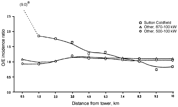
United Kingdom Studies. Dolk et al. (12) reported on leukemia incidence near the Sutton Coldfield radio and TV transmitters for the years 1974 to 1986. In addition, they studied adult leukemia incidence near 20 high power TV/frequency modulation (FM) transmitters in Great Britain (13).The Sutton Coldfield study examined data within a 10-km radius in 10 bands of increasing distance. The innermost area was within 2 km of the transmitter; adult leukemia relative risk (RR) was 1.83 (95% CI 1.22-2.74). Actually, one case lived within 0.5 km when 0.11 km could have been expected on the basis of cancer registry experience and the numbers of person-years of observation. While this results in an RR of 9, emphasizing location of a single case is likely to represent a poorly defined range of risk (Figure 1). There was a significant decline in risk with increased distance (p<0.001) from the transmitters. Expected numbers of leukemias in the 10-km zones near transmitters were calculated on the basis of national rates stratified by 5-year age groups, socioeconomic deprivation quintile, and region.
Figure 1. The 0/E leukemia incidence ratio by distance from TV and FM broadcast towers. The trends are shown for Sutton Coldfield and for two subsets of other such facilities in the United Kingdom for adults more than 15 years of age for the years 1974 to 1986. One subset is for facilities broadcasting TV in the range of 870 to 1000 kW and the second includes those with power from 500 to 1000 kW. The 0/E incidence ratio of 9.0 is based on a single case. Based on Dolk et al. (12,13).
In a second Dolk et al. (13) study the same procedures were used to evaluate risks surrounding 20 other broadcast facilities in the UK for the same period; 3305 cases were identified, with an overall observed-to-expected (O/E) ratio of 1.03 (95% CI 1.0-1.07). Decline in risk with distance was significant for all sites combined. Results in this study were similar to those of the Sutton Coldfield study (12). There was no significant excess risk for persons living within 2 km of the transmitters and excess risk was not greater than 15% in any distance band up to 10 km. However, the decline in risk for adult leukemia with distance from the transmitters was significant (p<0.05). Eight of the transmitters broadcast FM and three TV at power equivalent to transmission in the Sutton Coldfield study (12). One of the transmitters, Crystal Palace, was located in an unusually densely populated area and appeared to be associated with almost half the cases of leukemia. In the band between 2 and 3 km from the transmitter the adult leukemia O/E ratio was 1.33. Figure 1 shows some of these gradients for the Sutton Coldfield transmitters and for two other groups of stations, one with greater power than the other.
Rothman et al. (9) tabulated studies that might relate leukemia to
occupational or recreational exposures to RF radiation and studies that related
such exposures to brain malignancies. The risk ratios for leukemia were >1.0 for
19 studies and
![]() 1 for 7.
For brain tumors the RR was >1 for 9 studies, and
1 for 7.
For brain tumors the RR was >1 for 9 studies, and
![]() 1 for 4.
1 for 4.
Grayson (10) reported on brain cancer among U.S. Air Force personnel, and found that rank (socioeconomic factor) was the most important predictor. When this was taken into account, nonionizing radiation exposure was more important than ionizing radiation and microwave exposures more significant that low frequency exposures. The positive association for military rank had an OR of 3.30 (95% CI 1.99-5.45) for senior officers. For ionizing radiation, the association is negative. The military-rank-adjusted OR is significantly elevated for RF: 1.38 (95% CI 1.01-1.90), but not for ELF: 1.28 (CI 0.95-1.74).
Another study of military personnel and radiation exposures was that by Szmigielski (41), who examined cancer by site among Polish military personnel during the period 1970 to 1989. He found a relationship between exposures to high frequency (RF radiation) and cancer morbidity. About 3700 of the approximately 128,000 personnel were classified as exposed and data were tabulated for 12 types of cancer and four age groups. The overall cancer morbidity for exposed personnel was 119.1/100,000 per year compared on an age-adjusted basis to 57.6 in the nonexposed group. The greatest O/E ratios were found for chronic myelocytic leukemia, 13.9; myeloblastic leukemia, 8.62; and non-Hodgkin's lymphoma, 5.82.
A cluster of six cases of testicular cancer among traffic policemen using microwave generators suggests that microwave exposures can cause cancer of the testicle (42). Other epidemiologic studies of exposed military personnel point in the same direction (43,44).
Lin et al. (45) collected data on brain cancer deaths among white males for the state of Maryland, and examined occupations stated on the death certificates Included were 951 brain tumors, of which 370 were glioblastomas, 149 astrocytomas, and 432 had unspecified histology.
Fifty glioma and astrocytoma deaths among workers in occupations with a high probability of electrical exposures were matched by age with a sample of the population by age from the 1979 census. The expected number of such occupations in the general population was about one-third of that observed (18/50) for cases. A case reference study showed that the occupational category of electric or electronic engineer and technician had three times the number of cancer cases as the referent population (18 vs 6; p<0.05). When the specified occupations were ranked by definite, probable, or possible exposures to electromagnetic fields, the OR for astrocytoma and glioma were 2.15, 1.95, and 1.44, respectively.
Garland et al. (46) studied leukemia among occupational groups with potential electromagnetic field exposure in the U.S. Navy. Because they studied personnel who were hospitalized while on active duty, the study cannot include personnel with leukemia of substantial latency or those who were not hospitalized. In fact, one occupational group, electrician's mate(s), showed consistent excess of risk for leukemia.
Follow-up Study of 40,000 Korean War Naval Personnel. In the Robinette et al. (47) study, naval personnel were divided into occupational groups with low and high exposures by the occupational designator for the personnel. Within these two categories were three occupational classes, shown in Table 5.
Table 5. U.S. Naval personnel by occupational category during the Korean War and deaths by cause group. 1950 to 1974.
			         Low exposure                    High exposure    .
                           RM         RO        AE         ET         FT       AT
Number of persons          9253     10,116     1412      13,078     3298      3733
Total deaths                296       308       61         441        144      198
 From disease               161       165       22         199       81        77
 From malignant disease     39         47         8         65        16        27
 From malignancy of the lymphatic
  and hematopoietic systems   6        14         0         18         1        10

Abbreviations: ET, electronics technician(s); RO, radioman; RM. radarman. Data based on Robinette et al. (47).
Table 5 shows the occupational groups and numbers of cases. Table 6 gives rates for all deaths (per 1000) during the follow-up period of 1950 to 1974: rates for deaths attributable to disease, malignant disease, and malignancy of the lymphatic and hematopoietic systems. Death rates for the group with the highest exposure, aviation electronics technician(s) (AT), are significantly higher than those for the remaining men for all deaths, disease-related deaths, deaths from malignancy, and deaths from malignancy of the lymphatic and hematopoietic systems. Although it was true that this group had a higher mean age at onset of the follow-up study (23.4 years) than the average of the whole group (21.3) this mean age was younger than the average for aviation electrician's mate(s) AE (24.7), a category that showed no increase in deaths from any malignancy or from other diseases. The authors adjusted for age, but in doing so combined the AT group with the fire control technician(s) (FT) group, which had a low malignancy rate. These two groups, which were about the same size, had 10 and 1 cases of lymphopoeitic or hematological malignancies, respectively. For this population, compensated disability by body system is shown in Table 7 for the two high-exposure groups compared to the remainder of the population Both numbers and crude rates are given as well as the expected number of cases for the more exposed group based on the data for the remainder.
Table 6. U.S. Naval personnel by occupational category during the Korean War and crude death rates per 1000 by cause group, 1950 to 1974.
			         Low exposure                  High exposure        .
                           RM        RD         AE       ET         FT        AT
Number of persons         9253     10,116     1412     13,078      3298      3733
Total death rates           32.0      30.4      43.2       33.7      43.7      53.0
 From disease               17.4      16.3      15.6       15.2      24.6*     20.62*
 From malignant disease     4.21       4.65      5.66       4.97      4.85      7.23*
 From malignancy of the lymphatic
  and hematopoietic systems 0.65       1.38      0.00       1.38      0.3       2.68*

*  Significantly increased, p<0.05 compared to less-exposed groups. Data based on Robinette et al. (47).
   For occupational class definitions see Table 5 and text.
Table 7. Number of U.S. Naval personnel receiving Veterans Administration compensation in 1976, by diagnostic group, for two high-exposure groups (FT and AT) relative to the low-exposure groups exposed during the Korean War.
		         FT and AT                All others        FT and AT,
Diagnostic group      No       Rate/1000     No         Rate/1000   expected no
Musculoskeletal      119*       16.9         403          11.90        83.7
Special sense organs  42         6.0         152           4.49        31.6
Systematic conditions  5*        0.7           7           0.20         1.45
Respiratory           51*        7 3         171           5.05        35.5
Cardiovascular        47*        6.7         142           4.19        29.5
Digestive             55         7.8         229           6.76        47.6
Genitourinary         19         2.7          99           2.92        20.6
Hemic, lymphatic       3         0.4          10           0.30         2.08
Skin                  58         8.2         227           6.70        47.1
Endocrine             11         1.6          46           1.36         9.55
Neurologic            16         2.3          54           1.60        11.2
Nerves                 3         0.4          41           1.21         8.5
Epilepsies             0         -            16           0.47         3.32
Mental conditions     46         6.5         198           5.85        41.1
Other                  2         0.3          19           0.56         3.95
 Total diagnoses     477**      67.84         -           53.61       376.94
 Total populations  7031                   33,859

*  Significantly increased, p<0.05; ** significant, p<0.01. Data based on Robinette et al. (44).
Additional Studies of Cancer in Children and Others. Among the many tabulations from the Lilienfeld report (18), those for data about leukemia are shown in Table 8, based on data excerpted from the Lilienfeld report by Goldsmith (8). Although the numbers are small, there is significant excess for child dependents in both Moscow and other embassies, as well as an excess for employees and dependents in both locations. Estimated exposures at the Moscow embassy were from 5 to 18 µW/cm2.
Table 8. Leukemia among U.S. embassy employees and child dependents in Moscow and other Eastern European embassies.
	 	       Moscow embassy          Other embassies        Total
Population         Observed      Expected    Observed      Expected    0/E
Employees             2           0.8           3            1.7       5/2.5
Child dependents      2           0.5*          3            0.7*      5/1.2*
  Total               4           1.3*          6            2.4*     10/3.7*

*  Significantly elevated 0/E ratio, p<0.05. Based on table in Goldsmith (8).
Evidence of Other Health Effects
Lenticular Changes. Toncheva et al. (48) studied 87 persons working with radar and 150 eye-matched controls. The radar workers were divided into five risk groups according to frequencies of microwave exposure (200 KHz to 26 GHz) and power density (8 µW to 300 mW/cm2).
They found three specific radiation cataracts in persons working with extremely high microwave exposure. Lens changes were associated with level of exposure in different risk groups. Changes such as opacities and posterior polar defects are criteria for microwave exposure.
Nonspecific Disability. In their study of Korean War Veterans, Robinette et al. (47) obtained data for disability by body system in 1976. As noted in a previous analysis (8), the AT workers, those presumed to have received the most radiation exposures, were combined with the lesser-exposed FT to make what was designated the high-exposure group.
In the ten categories in Table 7 (categories with five or fewer cancer cases are not included) the high (FT+AT) group is higher than the remaining groups, with lower exposures in nine of ten body systems (significant by sign test at p<0.05).
The overall disability rate of 67.8/1000 is significantly greater than 43.1 by Poisson criteria. As is apparent from the combination of the two highest exposure job categories and the nature of the job classification procedure as described by the investigators, this analysis probably underestimates the effects of exposure.
Nonspecific Neurological and Sensitivity Reactions. Silverman (49) noted some nonspecific reactions to RF radiation, and a more recent review (50) brings these findings up to date. More research is needed to better define these reactions.
Available data suggest that RF radiation be considered a carcinogenic risk, a position already taken in an internal U.S. EPA document (51) in 1990 when there was much less evidence of the potential harmfulness of RF radiation.
Except for the Moscow staff, which includes both workers and dependents, most of the exposures studied are relevant to occupation. The most relevant to cases of community exposure risks today are those involving populations living near broadcast facilities. Cellular telephone users have not been exposed in definable numbers for a long enough time period for an adequate study to be made of cancer incidence.
However, interpretations must take into account the report of the Repacholi et al. study (52) of lymphoma-prone mice, who showed a doubling of the incidence of lymphoma over an 18-month period when exposed to modulated radiation similar to far-field cellular telephone exposures. This initial finding of experimental evidence of cancer from cellular-telephone-like exposures emphasizes the importance of examining epidemiologic evidence of such effects. Possibly the most suitable source for such data would be the more detailed study of exposures of military personnel or air traffic controllers who received definable exposures and have undergone a sufficient period of follow-up. Evaluation of such nonspecific symptoms as headache, sleep disturbances, and unfavorable reproductive outcomes of populations living near broadcast facilities should have priority for community studies.
The evidence may or may not justify more restrictive regulation of occupational exposure; for community exposures, however, the evidence justifies prudent avoidance (14,15). The concept has been presented by a group of Swedish government agencies in response to the evidence concerning ELF exposures. The plan is basically voluntary and stresses education about risks and economic analysis of uncertain risks and the possible costs of their avoidance.
Included among the actions to take under the rubric of prudent avoidance is epidemiologic monitoring (53), a system of standardized health status measurements of presumably reversible effects, which can, if unfavorable trends are discerned, become the basis for higher levels of population protection. The availability of a number of potentially reversible biologic responses makes this an unusually attractive possibility.
A second type of action is to provide realistic procedures to minimize the exposures. Shielding the head and face from exposures to the antennae of hand-held cellular telephones, and guidelines for keeping an adequate distance between broadcasting sources and civilian populations, are clearly indicated.
Further work is needed on the possibility of carcinogenicity in experimental systems of RF exposures. These systems should be separate from evaluations of ELF, which does not appear to have the same set of effects.
This review casts some doubt on efforts to distinguish ionizing from nonionizing radiation with respect to their health effects. It also raises doubt about the protective role of regulations based solely on the thermal effects of RF radiation, which is the basis for current standards.
There seems to be some evidence from the Moscow study and community studies in the vicinity of large FM and TV broadcasting facilities that exposures as low as 2 µW/cm2 may have long-term health effects.
A comprehensive and critical review of the epidemiologic data available on health risks from RF exposure should be carried out and the reasonable measures for avoidance of the identified risks should be described and evaluated.
References
1. Non-Ionizing Radiation: Proceedings of the 3rd International Non-Ionizing Radiation Workshop (Matthes R, ed), 22-26 April 1996, Baden (Vienna), Austria. Oberschleissheim, Germany:International Commission on Non-Ionizing Radiation Protection, 1996.
2. Feychting M, Ahlbom A. Childhood leukemia and residential exposure to weak extremely low frequency magnetic fields. Environ Health Perspect 103(Suppl 2):59-62 (1995).
3. International Commission on Non-Ionizing Radiation Protection, United Nations Environment Programme, WHO. Environmental Health Criteria 137: Electromagnetic Fields (300 Hz to 300 GHz). Geneva:World Health Organization, 1993.
4. United Nations Scientific Committee on the Effects of Atomic Radiation. Ionizing Radiation: Sources and Biological Effects. New York:United Nations, 1982.
5. Scotto J, Fears TR, Gori GB. Measurements of Ultraviolet Radiation in the United States and the Comparisons with Skin Cancer Data. DHEW (NIH) 76/1029. Washington:U.S. Department of Health, Education, and Welfare (National Cancer Institute), 1976.
6. Wertheimer N, Leeper E. Electrical wiring configurations and childhood cancer. Am J Epidemiol 109:273-284 (1979).
7. Ahlbom A. A review of the epidemiologic literature on magnetic fields and cancer. Scand J Work Environ Health 14:337-343 (1988).
8. Goldsmith JR. Epidemiological evidence of radiofrequency radiation (microwave) effects on health in military, broadcasting and occupational studies. Int J Occup Environ Health 1:47-57 (1995).
9. Rothman KJ, Chou CK, Funch DP, Dreyer NA. Assessment of cellular telephone and other radio frequency exposures for epidemiological research. Epidemiology 7:291-298 (1996).
10. Grayson JK. Radiation exposure, socio-economic status and brain tumor risk in the U.S. Air Force: a nested case-control Study. Am J Epidemiol 143:480-486 (1966).
11. Hocking B, Gordon I, Grain JL, Hatfield GE. Cancer incidence and mortality and proximity to TV towers. Med J Aust Assoc 165:601-605 (1996).
12. Dolk H, Shaddick G, Walls P, Grundy C, Thakrar B, Kleinschmitt I, Elliott P. Cancer incidence near radio and television transmitters in Great Britain. I: Sutton Coldfield transmitter. Am J Epidemiol 145:1-9 (1997).
13. Dolk H, Elliott P, Shaddick G, Walls P, Thakrar B. Cancer incidence near radio and television transmitters in Great Britain. Am J Epidemiol 145:10-17 (1997).
14. Aringer L. Unpublished data.
15. Swedish National Board of Occupational Safety and Health; National Board of Housing, Building and Planning; National Electrical Safety Board; National Board of Health and Welfare; Radiological Protection Institute. Low Frequency Electrical and Magnetic Fields: The Precautionary Principle for National Authorities: Guidance for Decision-Makers. ADI 478. Stockholm:Swedish National Board of Occupational Health, 1996.
16. Ouellet-Hellstrom R, Stewart WF. Miscarriages among female physiotherapists who report using radio- and microwave frequency electromagnetic radiation Am J Epidemiol 138:775-786 (1993).
17. Lilienfeld AM, Tonascia J, Tonascia S, Libauer CA, Cauthen GM. Foreign Service Health Status Study: Evaluation of Health Status of Foreign Service and Other Employees from Selected Eastern European Posts. Final Report Contract 6025-619073 (NTIS PB-288163). Washington:U.S. Department of State, 1978.
18. Jacobson G. Unpublished data.
19. Daily LE. A clinical study of the results of exposure of laboratory personnel to radar and high frequency radio. U.S. Naval Medical Bulletin 41:1052-1056 (1943). Cited in Steneck NH, Cook HJ, Vander AJ, Kane GL. Origins of U.S. safety standards for microwave radiation. Science 208:123-127 (1980).
20. Daily LE. A clinical study of the results of exposure of laboratory personnel to radar and high frequency radio. U.S. Naval Medical Bulletin 41:1052 (1943). Cited in Follis RH Jr. Studies on the biological effect of high frequency radiowaves (radar).Am J Physiol 147:281 (1946).
21. Barron CI, Love AA, Baraff AA. Physical evaluation of personnel exposed to microwave emanations. J Aviat Med 22:442-452 (1955). Cited in Steneck NH, Cook HJ, Vander AJ, Kane GL. Origins of U.S. safety standards for microwave radiation. Science 208:123-127 (1980).
22. Barron CI, Baraff FF. Medical considerations of exposure to microwaves (radar). JAMA 168:1194-1199 (1958). Cited in Steneck NH, Cook HJ, Vander AJ, Kane GL. Origins of U.S. safety standards for microwave radiation. Science 208:123-127 (1980).
23. Bach S. In: Proceedings of the 4th Tri-Service Conference on the Biological Hazards of Microwave Regulation, 16-18 August 1960, Griffis Air Force Base, Rome, New York. New York:Plenum, 1961;131-132. Cited in Steneck NH, Cook HJ, Vander AJ, Kane GL. Origins of U.S. safety standards for microwave radiation. Science 208:1230-1237 (1980).
24. Goldoni J. Hematological changes in peripheral blood of workers occupationally exposed to microwave radiation. Health Phys 58:205-207 (1990).
25. Goldoni J. Unpublished data.
26. Tonascia JA, Tonascia S. Unpublished data.
27. Vukelic M, Kontosic I, Jonjic A, Grubisic-Greblow H. Unpublished data.
28. Tornqvist S, Berggvist U, Hagman M, Knave B. Unpublished data.
29. Garaj-Vrohac V, Fucic A, Pevalek-Kozlina B. The rate of elimination of chromosomal aberrations after accidental exposure to microwaves. Bioelectrochem Bioenerg 30:319-325 (1993).
30. Balode Z. Assessment of radio-frequency electromagnetic radiation by the micronucleus test in bovine peripheral erythrocytes. Sci Total Environ 180:81-86 (1996).
31. Scarfi MR, Lioi MB, d'Ambrosio G, Massa R, Zeni O, Di Pietro R, Di Benadino D. Genotoxic effects of mitomycin-C and microwave radiation on bovine lymphocytes. Electro Magnetobiol 15:99-107 (1996).
32. Garaj-Vhrovac V, Horvat D, Koren Z. The effect of microwave radiation on the cell genome. Mutat Res 243:87-93 (1990).
33. Garaj-Vhrovac V, Horvat D, Koren Z. The relation between colony-forming ability, chromosome aberrations, and incidence of micronuclei in V79 Chinese hamster cells exposed to microwave radiation. Mutat Res 263:143-149 (1991).
34. Garaj-Vhrovac V, Fucic A, Horvat D.The correlation between the frequency of micronuclei and specific chromosome aberrations in human lymphocytes exposed to microwaves. Mutat Res 281:181-186 (1992).
35. d'Ambrosio G, Lioi MB, Massa R, Zeni O, Scarfi MR. Genotoxic effects of amplitude-modulated microwaves on human lymphocytes exposed in vitro under controlled conditions. Electro Magnetobiol 14:157-164 (1995).
36. Hagmar L, Brogger A, Hansteen IL, Heims S,Hogstedt B,Knudsen L, Lambert B, Linnainmaa K, Mitelman F, Hordensen I et al.Cancer risk in humans predicted by increased levels of chromosomal aberrations in lymphocytes. Cancer Res 54:2919-2922 (1994).
37. Akiyama M, Umeki S, Kusunoki Y, Kyoizumi S, Nakamura N, Mori T, Ishikawa Y, Yamakido M, Ohama K, Kodama T et al. Somatic cell mutations as a possible predictor of cancer. Health Phys 68:643-649 (1995).
38. Surveillance, Epidemiology, and End Results Program, National Cancer Institute. Cancer Incidence and Mortality in the United States, 1973-1981. Publ No 85-1837. Bethesda, MD:National Cancer Institute, 1984.
39. Anderson BS, Henderson AK. Unpublished data.
40. Maskarinec G, Cooper J. Investigation of a childhood leukemia cluster near low frequency radio towers in Hawaii [Abstract]. Am J Epidemiol 138:666 (1993).
41. Szmigielski S. Cancer morbidity in subjects occupationally exposed to high frequency (radiofrequency and microwave) electromagnetic radiation. Sci Total Environ 180:9-17 (1996).
42. Davis RL, Mostoff FK. Cluster of testicular cancer in police officers exposed to hand-held radar. Am J Ind Med 24:231-233 (1993).
43. Tarone RE, Hayes HM, Hoover RN, Rosenthal JF, Brown LM, Pottern LM, Javadpour N, O'Connell KJ, Stutzman RE. Service in Vietnam and risk of testicular cancer. J Natl Cancer Inst 83:1497-1499 (1991).
44. Bullman TA, Watanabe KK, Kang HK. Risk of testicular cancer associated with surrogate exposure measures of Agent Orange exposure among Vietnam veterans on the Agent Orange Registry. Ann Epidemiol 4:1-6 (1994).
45. Lin RS, Dischinger PC, Conde J, Farrell KP. Occupational exposure to electromagnetic fields and the occurrence of brain tumors: analysis of possible associations. J Occup Med 27:413-419 (1985).
46. Garland FC, Shaw E, Gorham ED, Garland CF, White MR, Sinsheimer PJ. Incidence of leukemia in occupations with potential electromagnetic field exposure in United States Navy Personnel. Am J Epidemiol 132:293-303 (1990).
47. Robinette CD, Silverman C, Jablon S. Effects upon health of occupational exposure to microwave radiation (radar). Am J Epidemiol 112:39-53 (1980).
48. Toncheva R, Zlateve B, Alexov D, Christova R. Unpublished data.
49. Silverman C. Nervous and behavioral effects of microwave radiations in humans. Am J Epidemiol 97:219-224 (1973).
50. Liakouris AGJ. Modulated Microwave Radiation from Soviet Medical Radar, Radiofrequency Sickness and the Lilienfeld Study. Carrboro, NC:Twin Streams Educational Center, 1996.
51. U.S. Environmental Protection Agency.Unpublished data. Cited in Sibbison JB. USA: Danger from electromagnetic fields. Lancet 336(8707):106 (1990).
52. Goldsmith JR, ed. Epidemiological Monitoring in Protection from Environmental Health Hazards. Sci Total Environ 32(3):211-363 (1984).
53. Repacholi MW, Basten A, Gebski V, Noonan D, Finnie J, Harris AW. Lymphomas in Eµ-Pim1 transgenic mice exposed to pulsed 900 MHz electromagnetic fields. Radiat Res 147:631-640 (1997).
Last Update: February 19, 1998