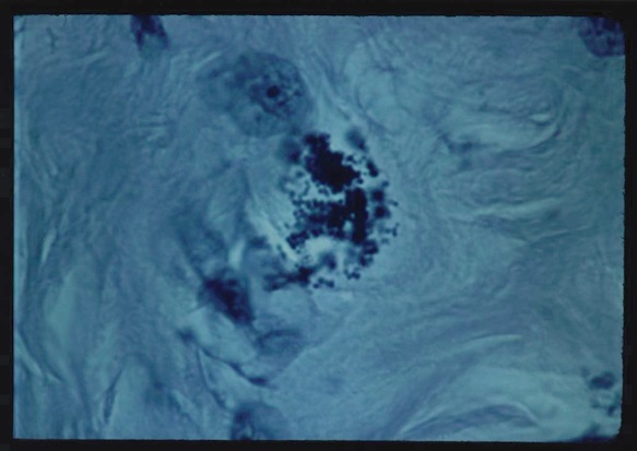
Figure 1. Histopathologic tissue section of original breast cancer showing grape-like clumps of variably-sized
Breast Cancer Is Caused By Pleomorphic Bacteria
© Copyright 2014
12-13-14
Bacteria were firmly rejected as a cause of cancer by medical science a hundred years ago. So why would any researcher claim that “cancer germs” cause breast cancer? It’s because pleomorphic bacteria can actually be seen in cancerous tissue; and a few physicians have been describing these unusual (and extremely controversial) bacteria since the late 19th century. Pleomorphic bacteria associated with cancer have more than one morphological form or appearance. When grown in lab culture and injected into mice they have been reported to increase the animals’ incidence of cancer. The microbe has been re-cultured from the animal cancer, this fulfilling “Koch’s postulates” — the proof required to establish a causative connection. For more details on pleomorphism in bacteria, consult the Wikipedia.
Since the “War on Cancer” in the 1960s, billions of dollars have been spent trying to find a viral (but not bacterial) cause of breast cancer. However, as of 2013 it now appears that viruses have finally been eliminated as a cause. Ka-Wei Tang, MD, one of the authors of a genetic analysis study of 4000 tumors, states: “In cancer research and treatment, there has been a lot of focus on associations that have not been proven, some of which have actually have been shown to be wrong. Researchers are starting to realize that we need truly unbiased methods to uncover meaningful associations.”
Unlike bacteria, viruses are too small to be viewed with the ordinary light microscope. But bacteria are large enough to be seen at the highest magnification (usually 1000 times, with an oiled lens) of the microscope.
William Russell (1852-1940) and "the parasite of cancer"
Near the close of the nineteenth century when major diseases like tuberculosis, leprosy, and syphilis were finally being widely accepted as bacterial infections, it was also thought that bacteria might also be implicated in cancer. On December 3, 1890, William Russell, a pathologist at the School of Medicine in Edinburgh, gave an address to the Pathological Society of London in which he outlined his microscopic findings of "a characteristic organism of cancer" that he observed in fuchsine-stained tissue sections from all forms of cancer he examined. He also noticed them in certain cases of tuberculosis, syphilis and skin infection.
The parasite was seen within the tissue cells and outside the cells (intracellular and extracellular). Their granular and round coccoid-shaped forms ranged in size from barely visible, up to "half again as large as a red blood corpuscle." The largest round forms suggested a fungal-like or yeast-like parasite. Russell provisionally classified the parasite as a possible "blastomycete" (a type of fungus); and called the tissue forms "fuchsine bodies" because of their bluish-red staining qualities.
At the time, it was thought that each species of microbe could only give rise to a single disease. In 1899, in yet another report on "The parasite of cancer" (The Lancet, April 29), Russell admitted he could sometimes detect cancer parasites in diseases other than cancer, and that this was indeed a "stumbling block." At this point a considerable number of scientists concluded that Russell bodies were merely the result of cellular degeneration of one kind or another. Furthermore, no consistent microbe was cultured from tumors; and attempts to induce cancer tumors in animals gave conflicting and often negative results. Russell was a pathologist, not a microbiologist, and he avoided controversies surrounding the cancer microbe theory. He simply concluded, "It seems almost needless to add that there remains abundant work to be done in this important and attractive field."
After three years' work at the New York State Pathological Laboratory of the University of Buffalo, Harvey Gaylord confirmed Russell's research in a 36-page report titled "The protozoon of cancer", published in May, 1901, in The American Journal of the Medical Sciences. Gaylord found Russell’s bodies in every cancer he examined, as well as large spherical bodies 2 to 3 times larger than red blood cells. But the most frequently seen round forms were the size of ordinary staphylococci.
Russell's “parasites” became widely known to pathologists as Russell bodies. They are currently considered to be “immunoglobulins” and non-microbial in origin. This report will suggest that pleomorphic Russell bodies actually represent extreme bacterial pleomorphism, a feature of so-called call wall-deficient bacteria described in recent decades. (For more on this, view ‘The Russell body: The forgotten clue to the bacterial cause of cancer’ (2003) and ‘The return of the cancer parasite’ (2011) online.
The heresy of the cancer microbe
The death kneel for a cancer germ resulted when famous pathologist James Ewing declared in 1919 that "Few competent observers consider it (the parasitic theory) as a possible explanation in cancer." Doctors eventually assessed cancer bacteria as laboratory contaminants or as secondary invader microbes that infect tissue after cancer has formed. Subsequently, few researchers dared to contradict Ewing by investigating bacteria in cancer.
Nevertheless, during the 1920s a few persistent physicians, such as pathologist John Nuzum of the University of Illinois College of Medicine; surgeon Michael Scott from Butte, Montana; and obstetrician James Young of Edinburgh, Scotland, published research showing that a particular type of bacteria was consistently observed and cultured in breast cancer. The pleomorphic germ defied the established laws of microbiology by its ability to change shape and form, depending on how it was cultured in the laboratory, the age of the culture, and the amount of oxygen supplied for growth.
At first, the germ in culture was barely visible as tiny round coccal forms. Later, these cocci might morph into rod-shaped bacteria, which could connect together to form filamentous chains resembling a fungus. Small cocci could also enlarge into yeast-like and fungal-like spore forms.
Nuzum grew his "micrococcus" from 38 of 41 early breast cancers, and from cancerous lymph nodes and metastatic tumors resulting from spread of the cancer to other parts of the body. With special stains he detected these small round coccoid forms within the breast cancer tumor cells. During his 6 years of intensive bacteriological study, he learned the microbe could pass through a filter designed to hold back bacteria, indicating that some forms of the microbe were as small as some viruses. Although Nuzum couldn't produce cancer tumors in mice, he was able to induce breast cancer tumors in 2 of 5 dogs injected with the microbe.
Young found his microbe in 16 cases of breast cancer, and in two mice with breast cancer. He identified "spore forms" and clumped "spore balls" in microscopic sections prepared from the mouse tumors. Scott described three stages in the life cycle of his pleomorphic bacteria —the rod forms, the spore or coccus-like forms, and larger spore-sacs resembling a fungus. He claimed to treat cancer patients with an effective antiserum against these microbes, and spent the rest of his life trying to alert colleagues to the infectious cause of cancer. But the antagonism to Scott's parasites and his antiserum was overwhelming, and he died a broken man. His sad story is told in The Cancer Conspiracy [1981] by Robert Netterberg and Robert Taylor.
The full papers of these men are seminal and valuable reading for anyone interested in the microbiology of breast cancer. They were retrieved for me by dedicated medical librarians who resurrected these totally forgotten scientific reports “long buried in the medical literature,” as my old dermatology profession used to say. It is unfortunate that they are not posted on the Internet by some well-funded breast cancer foundation seeking a cause and a cure.
Four women and their cancer bacteria discoveries
During the last half of the 20th century cancer microbe research was barely kept alive by a quartet of women, who were my friends but now have all passed away. The combined published research of Virginia Wuerthele-Caspe Livingston-Wheeler (a physician), Eleanor Alexander-Jackson (a microbiologist), Irene Diller (a cellular biologist) and Florence Seibert (a biochemist) provides indisputable evidence that bacteria are implicated in cancer.
Livingston independently discovered the cancer microbe in the late 1940s and never stopped promoting it until her death in 1990, at the age of 84. She stressed the pleomorphic microbe could be identified in tissue and in culture by use of the “acid-fast stain,” a traditional stain used to identify the rod forms of the tubercle bacillus and stains them a red color. She was greatly aided by TB microbe expert Alexander-Jackson, who supplied the bacteriologic expertise. Like previous researchers they confirmed the microbe was filterable and virus-like in some stages.
In 1965, they wrote: “This organism is a great simulator, whose various forms may resemble micrococci, diphtheroids, bacilli, fungi, viruses and host-cell inclusions. Yet if the developmental cycle of the organism is studied by following it through all its transitional stages, it can be identified as a single agent.” In 1974, the two women named their microbe "Progenitor cryptocides" (Greek for “hidden-killer”), causing an uproar among cancer experts, microbiologists, and the American Cancer Society, all of whom insisted such a perverse microbe did not exist!
In the 1950s Irene Diller of the Institute for Cancer Research at Fox Chase, Philadelphia, discovered fungus-like spicules emanating from cancer cells. Joining forces with the Livingston team, Diller worked with specially bred mice with a proven cancer incidence. By injecting them with bacteria cultured from breast cancer and other tumors, she was able to more than double the cancer incidence of the mice. When cancer tumors developed she successfully cultured the microbe from them — fulfilling Koch’s postulates. Diller also grew the microbe from the blood of cancer patients.
In the early 1960s Florence Seibert, noted biochemist and tuberculosis icon became so impressed with the three women’s research that she came out of retirement to help prove that bacteria caused cancer. Back in the 1920s Seibert devised a method to make intravenous transfusions safe by eliminating contaminating ubiquitous bacteria. Later, she perfected the skin test for tuberculosis that has been used worldwide ever since. In 1938, she was awarded the famed Trudeau Medal, the highest prize given to tuberculosis research.
Experiments conducted by Seibert and her team showed these TB-like cancer microbes were not laboratory contaminants because they were able to isolate bacteria from every tumor (and every leukemic blood) they studied. And when “immediate imprints” of fresh cancerous breast tissue were smeared onto a slide and stained, the coccus forms of the bacteria were easily identified. (See Figure 6A in her 1972 paper ‘Bacteria in Tumors’, written with Irene Diller and colleagues.)
In her autobiography, Pebbles on the Hill of a Scientist, published privately in 1968, she wrote: "One of the most interesting properties of these bacteria is their great pleomorphism. For example, they readily change their shape from round cocci, to elongated rods, and even to thread-like filaments depending upon what medium they grow on and how long they grow. This may be one of the reasons why they have been overlooked or considered to be heterogenous contaminants... And even more interesting than this is the fact that these bacteria have a filterable form in their life cycle; that is, that they can become so small that they pass through bacterial filters which hold back bacteria. This is what viruses do, and is one of the main criteria of a virus, separating them from bacteria. But the viruses also will not live on artificial media like these bacteria do. They need body tissue to grow on. Our filterable form, however, can be recovered again on ordinary artificial bacterial media and will grow on these. This should interest the virus workers very much and should cause them to ask themselves how many of the viruses may not be filterable forms of our bacteria."
Seibert's provocative papers, some published by the prestigious Annals of the New York Academy of Sciences, should have caused a stir. But with the quartet slowly closing in on the bacterial cause of cancer, funds from previous supporters (like the American Cancer Society) suddenly dried up. All cancer microbe researchers eventually discovered that studying cancer bacteria was the kiss of death as far as funding was concerned. And without adequate funding, cancer microbe research was made more difficult, if not impossible.
But coming from thirty years of TB research, Seibert knew that the discovery of a pleomorphic and intermittently acid-fast microbe in cancer was tremendously important. She fervently believed that knowledge of this microbe would be instrumental in developing a possible vaccine and more effective antibiotic therapy against cancer. In Pebbles she confided: "It is very difficult to understand the lack of interest, instead of great enthusiasm, that should follow such results, a lack of certainty not in the tradition of good science.” Like the other women, Seibert theorized the virus-like forms could disrupt and transform nuclear genetic cell material leading to malignant change. Even though cancer microbes might appear in the laboratory as simple and common microbes (like staphylococci and corynebacteria), their ability to infiltrate the cell nucleus meant they were far from harmless.
In 1990, at the age of 92, Florence B Seibert was inducted into the National Women's Hall of Fame. When she died the following year her passage was noted in Time and People magazines, and in major newspapers like The Los Angeles Times. All the obituaries mentioned her contributions to the safety of intravenous fluids and her great achievement with the TB skin test. But not a word was written about her cancer microbe research, to which she devoted the last years of her life.
Breast cancer bacteria in tissue and in culture
In early lab culture cancer bacteria may appear as ordinary staphylococci, particularly the coccus form of Staphylococcus epidermidis. The isolation of this common “normal” skin bacterium from any kind of cancer is generally considered insignificant. However, the demonstration of staph-like coccoid forms in breast cancer suggests this rejection may prove unwarranted. Furthermore, there is a little-known non-acid-fast coccus form of the TB bacillus (the mycococcus form), which is staphylococcal-like and indistinguishable from ordinary cocci. Using Google Scholar, one can view Anna Csillag’s entire 1964 paper ‘The mycococcus form of mycobacteria.’ British microbiologist and medical historian Milton Wainwright, in “Highly pleomorphic staphylococci as a cause of cancer” (2000), also provides scientific evidence to show why staph grown from cancer should be taken seriously and not be subject to automatic ridicule.
The following six microphotographs show the appearance of the intra- and extracellular, pleomorphic staphylococcus-like forms observed in acid-fast stained breast cancer tissue and in culture. All are derived from a 37 year-old woman, reported in 1981 by Cantwell and Kelso, diagnosed with “infiltrating ductal carcinoma of the breast” and treated with surgery and chemotherapy. Within months she developed metastatic skin tumors of her chest, and died the following year with widespread metastases. Acid-fast (red) and non-acid-fast (blue) rods forms (bacilli) were never seen. The size of the blue-stained round forms ranged from barely visible “granules,” but most of the coccoid forms were the size of ordinary staphylococci. Some attain the size of larger yeast-like and spore-like forms. (For more images of microbes in cancer tissue, Google my online postings: “Coccoid forms of bacteria and the cause of cancer,’ and ‘Bacteria cause cancer: The microscopic evidence’.)
Figure 1. Histopathologic tissue section of original breast cancer showing
grape-like clumps of variably-sized
non-acid-fast coccoid forms. Intensified Kinyoun's (acid-fast) stain, magnification x1000, in oil.
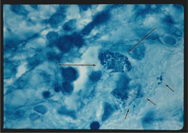
Figure 2. Breast cancer showing a clump of intracellular coccoid forms (long
arrows)
and scattered extracellular coccoid forms. Acid-fast stain, x1000, in oil.
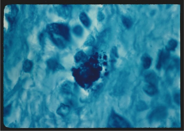
Figure 3. Breast cancer showing a tightly-packed intracellular clump of still
larger round coccoid forms. Acid-fast stain, x1000, in oil.
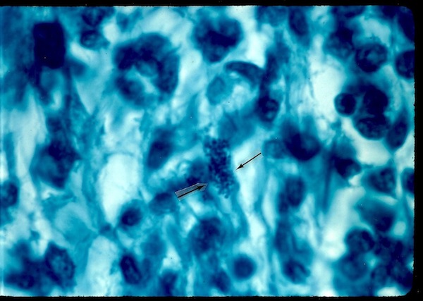
Figure 4. Breast cancer showing a focus of extracellular tiny "granular"
coccoid forms. Acid-fast stain, x1000, in oil.
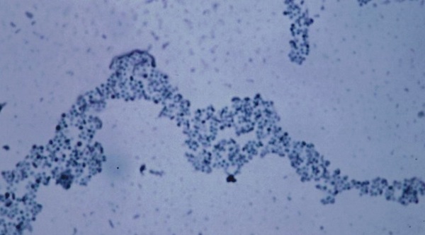
Figure 5. Staphylococcus epidermidis cultured from metastatic breast cancer
to the skin.
The cocci are not acid-fast. Compare the size and shape of these cocci to those seen in the original
breast cancer. Ziehl-Neelsen (acid-fast) stain, x1000, in oil.
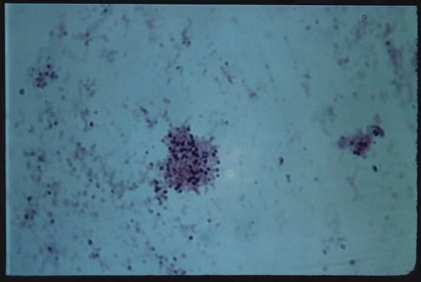
Figure 6. Staphylococcus epidermidis cultured from metastatic beast cancer to
the skin.
The staining quality of the cocci varies from Gram-positive (i.e. purple-stained) to pink. Gram stain, x1000, in oil.
Recent discoveries pertaining to cancer bacteria
In 2005 the Nobel Prize in Medicine was awarded to Australians Barry Marshall and Robin Warren for their discovery in the 1980s of the bacterium Helicobacter pylori and its role in gastritis and peptic ulcer disease. This infection can sometimes result in stomach cancer. The discovery proved that stomach ulcers were not due to diet, spicy food, alcohol, or psychiatric disorders, as previously believed. Physicians also previously believed that bacteria could not survive and flourish in the acid environment of the stomach. What was also essential in showing Helicobacter was the use of a special stain to identify these pleomorphic bacteria in stomach tissue. To also illustrate how one physician can be correct — and the rest wrong — is the fact that American pathologist A. Stone Freedberg [1908-2009] found similar bacteria in stomach ulcers decades before, in 1940. Unfortunately, he discontinued this research when doubting colleagues could not confirm his work and considered such an infection impossible.
In this new century the Human Microbiome Project has already revealed that 90% of the cells of the human body are not human cells. In actuality, the estimated 100 trillion cells are mostly bacterial in origin. Some writers now refer to us as “superorganisms.” Little is known about most of these germs and their affect on health and disease, especially diseases of old age and cancer. Livingston often said these body bacteria live peacefully within us in "symbiosis." However, when the immune system is weakened and/or when tissue is damaged, these bacteria proliferate and cause cellular inflammation. Inflammation is a precursor to the development of cancer; and cancer kills off one-third of the world’s population. According to the World Health Organization, one-third of the world’s population also has latent tuberculosis (TB).
The tragically forgotten cancer microbe
Why do so many physicians automatically reject cancer microbe research? Medical historian Lawrence Broxmeyer, MD, in ‘Cancer: A new perspective,’ (2011) posted online, believes James Ewing (now widely regarded as “the father of oncology”) condemned the research in 1919 because it conflicted with his radium interests and his financial investment in newly described radiation cancer therapy. In the 1950s prestigious pathologist Cornelius Rhoads also saw the research as a threat to his lucrative cancer chemotherapy interests. Broxmeyer’s controversial views are also expressed online in, ‘Is cancer just and incurable infectious disease?’ (2004) , as well as our co-authored ‘Is HIV a virus-like form of acid-fast tuberculosis type bacteria?’ (2008), posted on the www.joimr.org website.
In the years before her death Livingston was widely condemned as a quack by her colleagues. When she used an “autogenous” vaccine from the patient’s own cultured cancer bacteria to try to enhance the immune system response, she was highly criticized. (For more, read ‘Virginia Livingston: Cancer quack or medical genius?’) A damning report on her bacteria and vaccine entitled, ‘Autogenous vaccine: A defense against the bacterial organism that causes cancer, [2001]’ is presented online by Saul Green, PhD. Published by The Scientific Review of Alternative Medicine, he references 47 published papers, some of which are the same ones I use here to defend her work. However, he omits references to any of my 11 papers published in peer-reviewed journals illustrating cancer microbes in cancer tissue and in lab culture taken from patients with breast cancer, Kaposi’s sarcoma, and various forms of lymphoma. He also omits the formidable cancer research of Florence Seibert.
In my experience as a physician and octagenarian, I have learned that once doctors are carefully taught an important “fact” —such as “bacteria do not cause breast cancer” — it is difficult, if not impossible, for them to change their mind. I suppose it has to do with the security of being in sync with what your colleagues believe to be true and not making waves. With the demise of the “breast cancer virus” that so many oncologists believed in, perhaps we can again take a second look at cancer bacteria.
Sherlock Holmes once famously said to Doctor Watson: ‘You see but you do no observe.’ In this regard it took me a decade of observation of the microbe in various skin disease tissue to convince myself that Livingston and her women colleagues were correct. When I first met her in the mid-1960s, our mutual interest was primarily the very rare red-stained acid-fast TB-like rod forms we independently discovered in scleroderma tissue . For a long time I could “see,” but I did not carefully observe the other pleomorphic bacterial forms that were plentiful in scleroderma (especially the spectacular “large body” fungus-like forms I rarely observed). I was taught nothing about them in medical school and they weren’t found in medical textbooks. As a dermatologist in training, I couldn’t understand why expert microbiologists and pathologists did not report them, or provide a clear understanding of their origin. Finally, through painstaking observation, I developed the confidence to realize that microbes could indeed be seen in cancer, as other researchers had demonstrated years before.
For the prevention and treatment of cancer to advance significantly in this new century we have to understand clearly that “we” are microbes, and microbes are “us.” We are inseparable and we share an existential existence. Perhaps with that knowledge, we can begin to conquer cancer by maintaining and restoring the symbiosis between “them” and “us”.
SELECTED REFERENCES
Alexander-Jackson E: A specific type of microorganism isolated from animal and human cancer: Bacteriology of the organism.Growth 18:37-51, 1954.
Cantwell AR Jr, Kelso DW: Microbial findings in cancer of the breast and in their metastases to the skin. J Dermatol Surg Oncol7:483-491, 1981.
Cantwell AR Jr: The Cancer Microbe: The Hidden Killer in Cancer, AIDS, and Other Immune Diseases. Aries Rising Press, Los Angeles, 1990.
Diller IC: Growth and morphologic variability of pleomorphic, intermittently acid-fast organisms isolated from mouse, rat, and human malignant tissues. Growth 26:181-209, 1962.
Hess DJ: Can Bacteria Cause Cancer? Alternative Medicine Confronts Big Science. New York University Press, New York, 1997.
Livingston-Wheeler VWC, Addeo EG: The Conquest of Cancer. Franklin-Watts, New York, 1984.
Nuzum JW: A critical study of an organism associated with a transplantable carcinoma of the white mouse. Surg Gynecol Obstet33:167-175, 1921.
Nuzum JW: The experimental production of metastasizing carcinoma in the breast of the dog and primary epithelioma in man by repeated inoculation of a micrococcus isolated from human breast cancer. Surg Gynecol Obstet 11:343-352, 1925.
Scott MJ: The parasitic origin of carcinoma. Northwest Med 24:162-166, 1925.
Scott MJ: More about the parasitic origin of malignant epithelial growths. Northwest Med 25:492-498, 1925.
Seibert FB, Yeomans F, Baker JA, et al: Bacteria in tumors. Trans NY Acad Sci 34(6):504-533, 1972.
Wainwright M: Extreme pleomorphism and the bacterial life cycle: A forgotten controversy. Perspectives in Biology and Medicine 40:407-414, 1997.
Wainwright M: Highly pleomorphic staphylococci as a cause of cancer. Med Hypothesis 54:91-4, 2000.
Wuerthele Caspe (Livingston) V, Alexander-Jackson E, Anderson JA, et al: Cultural properties and pathogenicity of certain microorganisms obtained from various proliferative and neoplastic diseases. Amer J Med Sci 220:628-646, 1950.
Wuerthele-Caspe Livingston V, Alexander-Jackson E: An experimental biologic approach to the treatment of neoplastic disease. J Amer Med Women's Assn 20:858-866, 1965.
Wuerthele Caspe Livingston V, Livingston AM: Demonstration of Progenitor Cryptocides in the blood of patients with collagen and neoplastic diseases. Trans NY Acad Sci 34(5):433-453, 1972.
Wuerthele Caspe Livingston V, Livingston AM: Some cultural, immunological, and biochemical properties of Progenitor cryptocides. Trans NY Acad Sci 36(6):569-582, 1974.
Young J: Description of an organism obtained from carcinomatous growths. Edinburgh Med J (New Series) 27:212-221, 1921.
Young J: An address on a new outlook on cancer: Irritation and infection. Brit Med J, Jan 10, 1925, pp 60-64.