|
Fig. 1 |
Articles proving Vitamin C cures infections
In the treatment of hemorrhages and hemorrhagic tendencies of all kinds, vitamin C has proven quite effective, according to the works of BÖGER and MARTIN, BÖGER and SCHRÖDER, STEPP, SEDLACEK, ENGELKES, FREY, SCHLOESSMANN, SEYDERHELM among others. To what extent these can be compared as symptomatic of the most diverse illnesses, e.g. heavy infections, the “Werlhoff Illness” or intestinal hemorrhage in typhoid fever and paratyphus, icteric hemorrhages and finally blood diseases with a scurvey-like hemorrhagic tendency, and whether they are due essentially to vitamin C-deficiency, at present cannot be determined with confidence. Vitamin C exhibits numerous effects in the organism, of which the symptoms of clinical scurvy or prescurvy represent only one part of the deficiency symptoms; the effects of ascorbic acid on the cells, marrow and intermediate metabolism cannot be clearly separated.
In the treatment of blood illnesses and related conditions of other illnesses, vitamin C is recommended, according to recent works by ANDREU-URRA and REGLI, BÖGER und MARTIN, SCHADE, GRUNKE and OTTO, SCHNETZ, and, especially in leukemia, EUFINGER and GAETHGENS. Finally in this context the works of HAHN, CARRIÉ, SCHNEIDER, DEITEL, KUDLAC and STORM, VOGT, EDEL, among others, which report further on the efficacy of vitamin C doses on the side effects of x-ray treatment of malignant tumors, and likewise on the relatively frequently occurring “radiation leukopenia”.
On the basis of [our] own earlier observations during support of radiation treatment of malignant tumors by application of vitamin C, the impression was gained that vitamin C injections do not affect radiation leukopenia in any therapeutically usable measure; however, that it can favorably influence the [patient’s] general condition, and so be employed with advantage to support radiation treatment of malignant tumors. Success in recovery from radiation leukopenia is usually by injection of liver preparations, and in severe cases, by blood transfusions.
On the other hand vitamin C obviously has a favorable effect on “genuine leukopenia” or agranulocytosis. KALK reported recently on six patients with agranulocytosis, in which a reversal of the illness with an increase in granulated cells occurred through intravenous vitamin C injections.
The clinical and x-ray treatments of chronic leukemia must complement each other, and with the often years-long course of these illnesses, often enough the fate of the patient hangs upon which therapeutic measures are carried out. Vitamin C-only treatment is not recommended, apart from its very doubtful success, in not wanting to completely bypass 35 years of experience in the modern treatment of leukemia. For instance, clinical treatment trials conducted by H. E. BOCK, solely using vitamin C, were also unsuccessful. A comprehensive treatment of chronic leukemia in order to improve the general condition, as executed by us and others during the radiation treatment of malignant tumors, would seem to argue against it. At any rate, the effects of vitamin C during [treatment] are of use against the acutely aggravated tendency to hemorrhage often accompanying chronic leukemia.
In this context, the vitamin C expenditure of leukemia patients is to be reported briefly here. Balancing trials were made with 3 patients with myeloid and 3 patient with lymphatic leukemia, according to the loading method described elsewhere. Redoxon forte came to be used in treatment.
The method used was as follows: The vitamin C was administered intravenously, and in each case the excretion was monitored every 3 hours by titration of the fresh urine with Dichlorphenolindophenol by TILLMANNS [procedure]. The night urine collected in the morning at 8 o’clock was titrated. High doses were given that, as soon as the excretion in the urine increased, were then reduced, until after a day or two a larger excretion occured. We call the vitamin C quantity necessary for this the “current vitamin C deficit.” It is afterwards possible in most cases to maintain approximately the same urinary excretion of vitamin of C using smaller daily doses. This “running vitamin C deficit” determined thusly could, for other reasons, be resolved for only two of the patients. In regard to the particulars of this vitamin C loading method, one may refer to to the earlier paper. The following figures depict the course of the elimination and retention curves in leukemia.
Brief excerpts from the patient records are [included] to complement the curves charting the disease progression.
(Figures not included in this version)
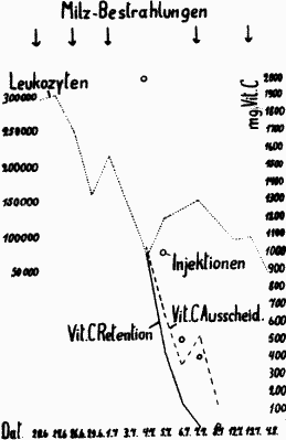
F. R., 55 years, butcher and innkeeper. Diagnosis: Chronic myeloid leukemia with distinct spleen enlargement. (see fig. 1). Family history unremarkable, moderate alcohol drinking. For approximately 4 months swollen feet in the evening, for 3 weeks the abdomen became distended with difficulty in breathing. Status: severely emaciated medium sized man, pale, cyanosis of the lips and ear lobes, moderate dyspnea. No lymph node enlargements. No cardiac enlargement. Blood pressure 130/75. Pulse a steady 120. Body: Spleen reaching up to the symphasis, the abdominal cavity filling out from the left to a handsbreadth over the center line to the right. Blood picture: Hb 67%, erythrocytes 3 million, leukocytes 293 000. Differentiation: unfragmented 18%, polymorphomuclears 27%, eos. 1½%, basoph. 3½%, lymphocytes 3% metamyelocytes 13½%, promyelocytes 3½%, myelocytes 23%, eosinophile myelocytes 2%, basophile myelocytes ½%, myeloblasts 3½%, normoblasts ½%, reticulum cells ½%. After treatment the spleen regressed by about half. Blood picture upon discharge: Hb 67% erythrocytes 3.6 million, leukocytes 50 000, differentiation: unfragmented 34%, polymorphomuclears 36%, eosinoph. 1%, basoph. 2%, myelocytes 12%, metamyelocytes 14%, eosinophile myelocytes 1%.
|
Fig. 1 |
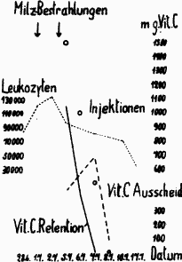
K. B., 55 years, housewife. Diagnosis: Chronic myeloid leukemia (see fig. 2). Family history unremarkable. 1927 Biliary colic. 1933 menopausal bleeding. Indeterminate, oppressive pain in the body for approximately ¾ year. Gum hemorrhages, fatigue. Findings: Pale woman in medium condition. Examination does not reveal special features, the spleen palpable, level with and under the rib elbow. Blood picture: Hb 82% Erythrocytes 4.8 Million, Leukocytes 83 000. Differentiation: Immature forms 5% Unfragmented 13% Polymorphomuclears 51% Eosinophile 3% Basophile 2% Lymphocytes 6% Myelocytes 18% Myeloblasts 2%. After treatment: Leukocytes 32 000, Immature forms 7% Unfragmented 18%, Polymorphomuclears 40%, Eosinophile l%, Basophile 2%, Lymphocytes 4%, Monocytes 8%, Myeloblasts 6%, Myelocytes 11%, plasma cells 3%. Improvement of the general condition, the spleen is no longer felt. Early release.
|
Fig. 2 |
With R., concerning advanced myeloid leukemia, the entire vitamin C retention amounted to 1840 mg. With B., concerning a moderately severe illness, which was so far improved by the treatment that the patient could perform her work as a housewife again, the vitamin C deficit amounted to 1470 mg. The vitamin C quantities of the “current deficit” do not represent particularly high values relative to those found with other illnesses. [We] also managed thus to reveal the deficit with several injections of Redoxon forte. A satisfactory improvement was achieved by the combined treatment in both cases.
The result of vitamin C loading differs under chronic lymphatic leukemias. Preliminary tests had shown that with these forms, especially with the presence of larger glandular nodes, a saturation of the “current deficit” was not achievable through moderate quantities of vitamin C. Therefore considerably higher vitamin C quantities had to be given.
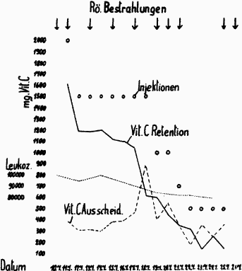
A. Sch., 58 years, sea grass processor*. Diagnosis: Chronic leukemic lymphatic Lymphadenosis with enlargement of numerous lymph nodes and spleen enlargement (see fig. 3). Family history unremarkable. For 20 years, enlarged glands at the neck, which however remained consistent. At the neck no operation scars. For 1 year, complaints of rheumatism. For ¼ year the lymph nodes at the neck enlarged, in addition to the appearance of lymph node enlargement in the arm pits and the groin flexure. In February 1939, initial hospital admission and irradiation of the tumors, the patient felt well and desired discharge. Second treatment in July 1939. General condition good, the tumors did not enlarge. Hilus enlargements are now determined on both sides. The spleen extends to a handsbreadth below the rib elbow. Blood picture: HB 85%, Erythrocytes 4 Million, leukocytes 100 000. Differentiation: Unfragmented 1%, Polymorphomuclears 3%, Eosinophile 1%, Lymphocytes 95%. This time irradiation of all the tumors was performed, especially on the hili and the spleen. The leukocytes dropped to 97 000.
|
Fig. 3 |
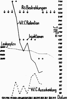
F. W., 55 years, wood cutter. Diagnosis: Chronic leukemic lymphatic lymphadenosis with substantial lymph node enlargements and large tumors in the abdominal cavity (see fig. 4).
|
Fig. 4 |
Family history unremarkable. Since 1935 feeling of abdomenal fullness, however no special complaints. Since August 1938, increase of the body circumference. Since September 1938, swelling of the neck. Shortness of breath. December 1938 admission to hospital. Lymphocytes 89 000. After irradiation 39 000. April through May 1939 2nd x-ray treatment. July August 1939 3rd. treatment. Blood picture: HB 90%, Erythrocytes 4 Million, leukocytes 62,000. Differentiation: Polymorphomuclears 6%, Lymphocytes 94%, blood corpuscle settling rate of 11 mm after WESTERGREEN. On both sides of the neck, in the armpits and groin flexure numerous to hen’s egg-sized lymph nodes. The abdomen is taut, the liver and spleen are undefinable opposite a lumpy tumor in the mid-abdomen. The hili are small apple-sized. In November—December 1939 4th treatment. The lymph nodes became substantially smaller, the leukocytes sank from 62 000 to 22 600.
The “current vitamin C deficit” with these two patients amounted to 11 820 mg and 11 240 mg. It is a matter of widespread leukemic lymphadenosis, with which the number of the white blood corpuscles stays within a moderate range. The “current deficit” with this and another patient amounted to about 200 mg.
Thus it was shown that the vitamin C deficit with leukemic lymphatic lymphadenosis was larger by several times relative to myeloid leukemia. Earlier vitamin C-balance investigations with Lymphogranulomatosis and tumors with metastases, which were similarly conducted regarding the spread of neoplasms, but not regarding the description of the clinical disease’s severity, found a vitamin C deficit of 1000-3000 mg, and in very serious cases of 5000 mg. It is shown that with leukemic lymphadenosis, even if the illness is not yet very life-threatening, a substantial vitamin C deficit is already present. Apart from the reasons already stated, this demonstrable deficit warrants the application of vitamin C, particularly in the treatment of the lymphatic forms of chronic leukemia.
As to the cause of the vitamin C deficit, which is found to this extent only with scurvy and with other very severe acute illnesses, not much can be stated at present. With the investigated myeloid leukemias, the determined current deficit of 1470 mg and 1840 mg corresponds to the deficit present with other types of similarly severe illnesses. The values determined with the lymphatic forms are far higher in comparison. Since a saturation of the deficit can be achieved up to certain degree, even if x-ray irradiations are not performed, the conclusion is justified that hypovitaminosis-C is also present here. That the strongly active tumor tissue growth present in lymphatic lymphadenosis leads to an increased vitamin C consumption is to be assumed. However, after the histochemical investigations of TONUTTI, in tissues exhibiting increased activity and with a sufficient supply of vitamin C, a plentiful accumulation of the same is found. Under these circumstances the administration of vitamin C would cover this increased consumption. However, what is not yet finally decided is the important question of whether covering the demonstrated vitamin C deficit increases the activity of the storing tissue, thereby aggravating the disease process, or whether supplying the missing vitamin C quantity affects the illness favorably. Relative to leukemics not treated with vitamin C, the continued progression [of disease] showed, at re-examinations and later treatment, no difference and particularly no increase in expected relapses.
The treatment of chronic myeloid and lymphatic leukemias only with vitamin C does not lead to satisfying success. Nevertheless vitamin C favorably affects the general state, and is indicated in hemorrhagic tendencies. With myeloid leukemias the occasionally saturatable “current vitamin C deficit” amounted to 1000-2000 mg. With leukemic lymphadenosis the “current deficit” was substantially higher, amounting in moderately severe cases to 10 000 to 12 000 mg.; with these, the “current vitamin C deficit” amounted to about 200-300 mg. The cause for this vitamin C deficit may be attributed after TONUTTI to increased cell activity.
(Anschr. des Verf.: z. Z. Wildbad i. Schw., Reservelazarett)
From Deutsche Medizinische Wochenschrift, April 5, 1940, Number 14, pp. 369-372
Translator’s notes:
The translator wishes to give sincere thanks to the contributers at sci.lang.translation for their tips and observations.
HTML Revised
04 November, 2015.
Formatting © 1999 AscorbateWeb
Translation © 1999 by Alexander Stoll