Vaccines, Neurodevelopment and Autism Spectrum Disorders
The Danger of Excessive Vaccination During Brain Development: The Case for a Link to Autism Spectrum Disorders
March 12, 2008
The Autism Disaster: Is it Man Made?
Autoimmunity and Vaccinations
Immune Suppression By Live Virus Containing Vaccines
The Autistic Prone Child
Seizures and Autism
Human Brain Development is Different
What About the Adjuvants Used in Vaccines?
The Role of Mercury in Developmental Brain Damage
Why Males Are Affected More Often
The Role of the Leaky Gut Phenomenon and Food Intolerances.
Conclusion
In 1976, children received 10 vaccines before attending school. Today they will
receive over 36 injections. The American Academy of Pediatrics and the Center
for Disease Control assured parents that it was safe to not only give these
vaccines, but that they could be given at one time with complete safety. Is this
true? Or are we being lied to on a grand scale?
The medical establishment has created a set of terms, which they use constantly
to boost their egos and firm up their authority as the unique holders of medical
wisdom–the mantra is “evidence-based medicine", as if everything outside
their anointing touch is bogus and suspect. A careful examination of many of the
accepted treatments reveals that most have little or no scientific
“evidence-based” data to support it. One often repeated study found that almost
80% of medical practice had no scientific backing.
This is not to say that medical practice should be purely based on pure and
applied science, as understood in the fields of physics and chemistry. Medicine,
as pointed out by many of the great men of medicine, is an art. For a discussion
on the proper role of medicine I refer the reader to my paper titled –Regimentation
in Medicine and the Death of Creativity– on my website (www.russellblaylockmd.com).
Most men of medicine recognize that some things are obvious without a placebo
controlled, double-blind, randomized study. For example, there has never been
such a study to see if smashing your finger with a hammer will be painful, but
we accept it without such pristine evidence. The same is true with removing
brain tumors or sewing up severe lacerations.
I find it interesting that there exist an incredible double standard when it
comes to our evidence versus theirs. The proponents of vaccination
safety can just say they are safe, without any supporting evidence what-so-ever,
and it is to be accepted without question. They can announce that mercury is not
only safe, but that it seems to actually increase the IQ, and we are to accept
it. They can proclaim thimerosal safe to use in vaccines without their having
ever been a single study on its safety in over 60 years of use, and we are to
accept it.
Yet, let me, or anyone else, suggest that excessive vaccination can increase the
risk of not only autism, but also schizophrenia and neurodegenerative diseases,
and they will scream like banshees –Where is the evidence? Where is the
evidence? When we produce study after study, they always proclaim them to be
insufficient evidence or unacceptable studies. More often than not, they just
completely ignore the evidence. This is despite the fact that we produce dozens
or even hundreds of studies that not only demonstrate the link clinically and
scientifically, but also clearly show the mechanism by which the damage is being
done –even on a molecular level. These include cell culture studies, mixed cell
cultures, organotypic tissue studies, in vivo animal studies using
multiple species and even human studies. To the defenders of vaccine safety-our
evidence is never sufficient and, if we face reality –never will be.
When I was in medical school, there was no proof that cigarette smoking cause
lung cancer. The connection was as obvious as the layman’s observation that
smashing your finger with a hammer would cause pain and even the town drunk knew
it was true, but to the medical elite –there was no proof.
No one had ever produced lung cancer in animals by exposing them to cigarette
smoke. In fact, my pathology professor, Dr. Jack Strong, had trained monkeys to
chain smoke, and after years of smoking none developed lung cancer. Yet, he was
convinced that smoking caused lung cancer. Dr. Alton Oschner, founder of the
famed Oschner Clinic in New Orleans, led the charge in proclaiming the link
between cigarette smoking and lung cancer. It took almost another decade before
the medical elite was willing to admit that smoking caused most cases of lung
cancer.
Almost 30 years passed from the time some iconoclastic men of medicine tried to
convince the medical establishment that smoking caused most cases of lung cancer
until it was generally accepted. The questions that needs to be asked is –How
many people died of lung cancer, the most prevalent cause of cancer death in the
United States, during this time? Data from the National Cancer Institute
estimated that in the year 2004, 157,000 people died of lung cancer. If 80% were
secondary to smoking that would be 125,000 dead. Over a ten-year period that
would be over one million dead and over 30 years almost 4 million people who
died from a preventable cause of death that at the time was still being hotly
debated by the medical purist. Lung cancer death rates were actually higher
during that time period.
So we see that questions of medical importance that are nick picked to death on
points of scientific purity can cost a lot of lives –millions of lives. There
are over one million children and even adults with autism and the numbers
continue to grow. This is a medial disaster of monumental proportions. The link
to the vaccine program is scientifically and logically compelling but these same
medical elitists refuse to listen.
Like smoking and lung cancer, we have enough proof today to call a halt to the
present excessive vaccine program and ban any level of mercury in vaccines. In
1983, before the autism epidemic began, children received 10 vaccinations before
attending school and the autism incidence was 1 in 10,000. Today they are
receiving 23 vaccines before age 2 years and 36 by the time they attend school
and the autism rate is now 1 in 150 births. Medical “experts” have provided no
other explanation for this dramatic and sudden rise in autism cases, despite a
draconian effort to find one.
They attempted to say it was genetic, but geneticists were quick to respond that
genetic disorders do not suddenly increase in such astronomical proportions.
They then said it was because of better diagnosis, despite the fact that the
diagnosis is obvious in virtually every case and that the criteria officially
accepted for diagnosis has become more restrictive not less.
When trapped by a lack of evidence, defenders of a nefarious position resort to
their old standby –the epidemiological study. Statisticians will tell you that
the least reliable type of study is an epidemiological study because it is easy
to manipulate the data so that the study tells you anything you wish it to.
Every defense offered by vaccine defenders is based on such studies and never
the actual science. Then they announce that the issue is settled and no further
studies need be done. After the media has been informed that the issue has been
settled, those who continue to present the evidence are considered kooks and the
great unwashed ignorant.
The Autism Disaster: Is it Man Made?
Today, specialists speak of the autism spectrum disorders (ASD), which include a
number of related neurodevelopmental disorders such as classical autism, Rett’s
syndrome, Asperger’s syndrome, childhood disintegrative disorder (CDD) and
pervasive developmental disorders not otherwise specified (PDD-NOS). I have
noticed over the years that when specialists know very little about a disorder
they spend an inordinate amount of time naming and sub-classifying it
–periodically. In addition they go to great lengths to define characteristics
and symptoms of the disorder that must be present to meet the criteria of
classification. Those who fail to meet these criteria are dispensed with into
another dimension, that is, they are ignored.
In the early 1980s, the incidence of autism was 1 in10,000 births. By 2005, the
incidence had leaped to 1 in 250 births and today it is 1 in 150 births and
still climbing. One of the strongest links to this terrible set of disorders was
a drastic change in the vaccine programs of the United States and many other
countries, which included a dramatic increase in the number of vaccines being
given at a very early age. No other explanation has been forthcoming from the
medical elite.
In this paper I shall present evidence, some of which has not been adequately
discussed, that provides strong evidence for a connection between excessive
vaccination and neurodevelopmental disorders. In a paper I wrote in 2003, I
stated that removing the mercury from vaccines would help relieve the problem,
but it would not eliminate it. This was based on a number of studies in the
neuroscience literature that indicated that excessive and especially repeated
immune stimulation could result in severe disruption of brain development and
even neurodegeneration.
In this paper and a follow-up paper, I attributed the central mechanism to
excessive and prolonged microglial activation with an interaction between
inflammatory cytokines and glutamate receptor subtypes. The Vargas et al study,
published two years later in 2005, strongly supported this hypothesis, with the
finding of elevated inflammatory cytokines as well as the presence of extensive,
widespread activated microglia and astrocytes in examined autistic brains from
age 5 years to 44 years of age. This indicated that the brain’s immune
activation persisted for decades. Recent research indicates that this phenomenon
is not that uncommon and can be reproduced in the laboratory using a variety of
immune stimulating agents and neurotoxins, including mercury and aluminum.
Autoimmunity and Vaccinations
A number of studies have suggested a link between autoimmune disorders and
autism risk. Support comes from studies showing an increased risk of ASD in
children of mothers with autoimmune disorders.1-3 Yet, not all studies agree,
since at least one carefully done study found no strong link.4
Other more carefully done studies provided evidence suggesting some link. For
example, in one study serum from a mother with an autistic child was found to
bind immunologically with specific brain cells (Purkinje cells).5 When this
serum was injected into pregnant mice, their babies demonstrated neurological
changes suggestive of autistic behavior, indicating a transfer of the
autoantibodies into the developing baby mouse.
A number of studies have found autoantibodies in a significantly higher number
of autistic children to various brain structures, such as serotonin receptors,
myelin basic protein, neuron axon filament protein, nerve growth factor and
cerebellar neurofilaments.6-10 It should be understood that these autoantibodies
are not found in all cases and that they may develop as a result of the damage
caused by the disease itself, rather than causing the disease. For example, we
know that after a stroke or head injury a substantial number of people will
develop autoantibodies to brain proteins. Never the less, the autoantibodies can
worsen the damage and prolong the damaging pathology.
It has also been demonstrated that methylmercury (from fish) and ethylmercury
(in thimerosal) are both powerful immunosuppressants and are associated with a
high incidence of autoimmunity.11 In this study, researchers found that unlike
methylmercury, thimerosal (ethylmercury) initially caused immune suppression and
then strong TH2-induced autoimmunity. They attributed this to the higher
conversion of ethylmercury to ionic mercury (Hg+) than seen with methylmercury.
In fact, one study found that strains of mice highly susceptible to developing
autoimmune diseases were sensitive to the ASD-like behavioral effects upon
mercury exposure, whereas mouse strains genetically not susceptible to
autoimmunity do not develop ASD behaviors.12 It is obvious from the extremely
high incidence of ASD that these autoimmune-related genes are very common, but
they remain silent until triggered by vaccines or other environmental toxins.
Immunologists have now concluded that autoimmune disorders are not the result of
excessive activation of a normal immune system, but rather activation of a
dysfunctional immune system. The question remains- what is causing such
widespread immune dysfunction among our population? Studies have shown that the
number of autoimmune diseases has increased over the past 30 years, with asthma,
type 1 diabetes and eczema rates increasing over two fold. There is also
compelling evidence to indicate that certain vaccinations are associated with
these autoimmune-related conditions.13,14
A compelling number of studies have shown an increase incidence of autoimmune
reactions in children with the autism spectrum disorders (ASD), especially
involving measles antigens, milk antigens and antibodies to gliadin and
gluten.15-17 Some of these have been shown to cross-react with brain-derived
proteins as well, especially those in the cerebellum, a major structure affected
in these disorders.18
Recently, neuroscientists have shown that much of the damage done in cases of
autoimmunity is not due to direct immune reactions with brain structures, but
rather results from the release of storms of free radicals and lipid
peroxidation products during the immune reaction, something I call a “hand
grenade in a shopping mall effect”. If you use a hand grenade to target a single
person in a crowd you will not only kill and injure the intended target, but all
of the bystanders as well.
Neuroscientists P.L. McGeer and E.G. McGeer have named this effect bystander
damage.19 The immune attack caused by the autoimmune reaction in the
autistic person’s brain damages a number of surrounding structures, especially
brain connections called dendrites and synapses. Subsequent studies have
confirmed that bystander damage is the most destructive reaction of
autoimmunity.
Some studies, as referred to above, have shown that autism is much more common
in families with a hereditary tendency for autoimmune diseases, which makes
sense because they will have dysfunctional immune systems. There is also
compelling evidence that vaccines themselves can damage the immune system of
immature animals, leading to a higher incidence of autoimmunity and abnormal
brain development.20-24 Mercury, even in small concentrations, is also known to
induce autoimmunity in a high percentage of those exposed.11
Ironically, things that suppress a portion of the immune system, usually
cellular type immunity, increase the likelihood of autoimmunity. Immunologists
speak about a Th1 to Th2 shift and vice versa. This can occur with exposure to
mercury as well as in response to vaccination.25 A great number of autoimmune
diseases are associated with a Th2 shift.
The immune system is a very complex system, which at birth is incompletely
formed. This means, and has been confirmed in animal and human studies, that
immune reactions to vaccinations differ at different ages, so that small babies
have a different reaction than adults. This has been shown with the hepatitis B
vaccine now given to newborns. The rate of maturation of the immune system also
differs considerably among babies and children, meaning we cannot say what
effect will occur in all children. There are a great many variables, including
diet.
The immune system’s reaction to infection and immunization can be quite
different. Normally the immune system relies on a shifting of T-lymphocyte
function to determine which is better for the particular situation.26 The
T-helper lymphocytes (Th) can exist as either Th1, Th0, or Th2 forms. When no
infection is occurring, the system is in the Th0 mode (an uncommitted phase). If
a virus invades, it quickly switches to the Th1 phase, which allows immune cells
to secrete a group of cytokines that kill viruses. It also activates immune
lymphocytes that kill viruses and bacteria. At other times, the immune system
needs a whole different set of immune signals and cells, which are supplied by
the Th2 phase. The Th2 phase favors the production of antibodies, mainly
supplied by B-cells, but in general they reduce immune reactions.
Infants are stuck in the Th2 mode during intrauterine life, so as to prevent
being immunologically rejected by the mother during pregnancy (much like
transplant rejection), since the baby is seen as a foreign body to the mother’s
immune system. Upon birth, the baby remains in a Th2 mode, but has a limited
ability to switch to the Th1 defensive mode if the need arises, say from an
infection. Months later the baby switches to the adult Th1 mode. If the baby’s
immune system remains in a Th2 mode, it has a high risk of developing an
autoimmune disorder, such as eczema, asthma or other allergies.
Presently, vaccine authorities recommend every baby be vaccinated with the
Hepatitis B vaccine at birth. But, is this safe? A recent study looked at the
immune reaction in newborn infants up to the age of one year who had received
the HepB vaccine to see if their immune reaction differed from adults getting
the same vaccine.27 What they found was that the infant, even after age one
year, did react differently. Their antibody levels were substantially higher
than adults (3-fold higher) and it remained higher throughout the study. In
essence, they found that the babies responded to the vaccine by having an
intense Th2 response that persisted long after it should have disappeared, a
completely abnormal response.
Autistic children have been described as having a Th2 predominance, which would
explain their propensity to developing autoimmune diseases and being more
susceptible to infections early in life.20,28-30 Elevated proinflammatory
cytokines, particularly TNF-, have been described in studies of the cytokine
profile in autistic children. As we shall see later, an excess production of
B-cell cytokines and suppression of T-lymphocyte TH1 activity, as seen in
autism, is associated with a high incidence of neurological damage by
excitotoxins.
Several things about these immune responses are important to all parents,
including effects of such immune overstimulation during pregnancy. For example,
it has been shown that excess immune stimulation, as occurs with vaccination,
can significantly increase the risk of a pregnant woman having a child with
autism or schizophrenia later in life, depending on when the vaccine is
given.31.32 In addition, persistent Th2 responses caused by the HepB vaccine
puts your child at a great risk of developing an autoimmune disorder and
impairing your baby’s ability to fight off infections. This means that
immediately after birth this vaccine has put your child at a greater risk of all
childhood related infections, including H. Influenza meningitis, meningiococcal
meningitis, rotavirus, measles, chickenpox, etc. Not only that, but numerous
studies have shown that such immune suppression greatly increases the number of
severe complications associated with these infections, which means that should
your child be exposed to measles or chickenpox they are more likely to suffer
neurological damage, seizures or other systemic disorders.12,33,34 When this
occurs, rather than admit that the science indicates that the vaccine program is
the cause of the complications and deaths, the vaccine proponents scream that it
demonstrates again the need for greater efforts to vaccinate our children.
Immune Suppression By Live Virus Containing Vaccines
It is also known that certain viruses powerfully suppress immunity, such as the
measles virus.35 The MMR vaccine contains live measles viruses and recent
studies have shown that immune suppression after vaccination with this virus
suppresses immunity in a profound way that last as long as six months.36-41 In
fact, the CDC recommends separating this vaccine from other live virus vaccines
to prevent viral overgrowth (Yet, they combine it with two other live
viruses-rubella and mumps viruses).
Yet, they never address the obvious question –wouldn’t this vaccine also make
the child more susceptible to other naturally occurring infections such as
hemophilus B influenza meningitis, meningococcal meningitis, persistent measles
infection, influenza infection and even chickenpox? This has been strongly
suggested by a number of studies.42 Not only would they be more susceptible, but
severe complications and even death would be more common as well.
When death and severe complications occur due to these infections, pediatricians,
the CDC and the American Academy of Pediatrics use this as a justification for
more vaccines, never admitting that the increase incidence of these infections
and complications was caused by their previous vaccine recommendations.
This risk is especially high in families with a number of other children in the
household or in children in day care centers. With a prolonged suppressed immune
system, exposure to other sick children would put this child at a high risk of
contracting the infection and of having complications or dying from the
infection as stated.
Studies have also shown that vaccines that cover only a few strains of a virus
or bacteria that naturally have a great number of strains (some have over a
hundred strains), can cause a shift in strain dominance so that the strain not
included in the vaccine then becomes the dominant disease causing strain. We see
this with the meningiococcal and pneumococcal vaccines.43-45 This is discussed
in the scientific literature but the public is never informed. Most
pediatricians are completely unaware of this.
When combined with mercury, which is also an immune suppressing substance, the
effect is compounded. Fluoroaluminum, formed in fluoridated drinking water, also
interferes with immune function, as do many insecticides and herbicides used
around the home.46
Often forgotten, is the substantial evidence that omega-6 oils powerfully induce
inflammation and immune suppression when consumed in large amounts. Those eating
a Western diet are consuming 50-fold higher amounts of this type of oil (called
linoleic acid) than needed for health. These oils include corn, safflower,
sunflower, canola, peanut and soybean oils. So, we see that the average child is
exposed to a number of substances in their food and environment that can also
alter immunity, making them not only more susceptible to natural infection, but
also to vaccine complications.
In essence, by overvaccinating our children, public health officials are
weakening their immune system, making them more susceptible to a number of
infections and less able to combat the infections. This gives them an endless
source of “horror stories” to justify even more vaccines. Remember also that
mercury is an immune suppressant, that both from vaccines and seafood
contamination.
One can see that a pregnant mother having dental amalgam fillings who eats a
diet high in methylmercury-containing seafood and living in an area with high
atmospheric mercury, such as West Texas, would be at a greater risk of having an
autistic child than one not exposed to these other sources of mercury. These
differences in environmental mercury exposure are never considered by those
insisting all children have the same vaccines, including mercury-containing
vaccines such as the flu vaccine.
The Autistic Prone Child
What is becoming obvious is that certain children are at a higher risk of
developing autism than others, for a variety of reasons. It is also obvious that
these newborns and small children develop infections at a higher rate than less
vulnerable children. This may be because of a developmental immune deficiency,
which can affect only a portion of the immune system and so be easily missed by
their pediatrician. Indeed, it has been noted that a great number of cases of
childhood immune deficiencies are missed by practicing pediatricians, especially
the more subtle cases, which may make up the majority of ASD-prone children.
For example, many physicians treating autistic children have noted a high
incidence of ear infections. These are treated with broad-spectrum antibiotics,
which often lead to a high incidence of Candida overgrowth in the child’s body.
Both infections will prime the microglia in the child’s brain –which is the
brain’s specific resident immune cell. This priming effect shifts these normally
resting microglia immune cells into overdrive.47 If stimulated again within
weeks or even months, they generate extremely high levels of free radicals,
lipid peroxidation products, inflammatory cytokines and two excitotoxins
glutamate and quinolinic acid.48 Studies have shown that this is the major
mechanism for both viral and vaccine-related brain injury.
The high incidence of infection in these children indicates the possibility of
preexisting immune system dysfunction. As stated, this also increases the risk
of an autoimmune reaction. The stage is then set for the autism cascade to
develop and this can be triggered by early vaccination or a recurrent infection.
Remember, the microglia have been primed, either by a natural infection or an
earlier vaccination (such as the hepatitis B vaccine given soon after birth).
The vaccine is different from a natural infection in that the vaccine produces
brain immune stimulation for very prolonged periods.
It has been proven, in both animal studies and human studies, that systemic
infections or immune activation by vaccines, rapidly activate the brain’s
microglial system and can do so for prolonged periods.49-53 Once the primed
microglia are reactivated by the subsequent vaccination or infection, the
microglia activate fully and pour out their destructive elements as discussed
above.
With a natural infection, the immune system quickly clears the infection and
then shuts off the immune activation, thus allowing repair of what damage was
done. This shutting down of the microglia is very important. There is evidence
that with repeated and excessive vaccine-triggered immune stimulation, the
microglia do not shut down.47 This is what was found in the Vargas et al study,
in which they examined the brains of 11 autistics from age 5 years to 44 years
of age dying without active infectious diseases as compared to age matched
controls.54 That is, they found widespread activation of inflammatory cells (microglia
and astrocytes) in the brains of the autistic patients. This explains the
widespread brain damage seen in all autism cases.
This study was one of the most carefully conducted, extensive examinations of
the immune reactions in the autistic brain ever done and involved
immunocytochemistry, cytokine protein assays and enzyme-linked immunoascorbant
assays of the brain tissue. They also performed similar assays of spinal fluid
from an additional six living autistic patients, which confirmed the intense
immune activation and inflammation.
The average child receiving all of the recommended vaccines will have some 23
inoculations by age two years and 36 by the time they enter school. Most of
these will be spaced within one month of each other, which means the priming and
activation cycle of the microglia will be continuous. In addition, should
they follow the new CDC recommendation, they will receive the flu vaccine every
year starting at age 6 month through age 18 years. These vaccines contain a full
dose of thimerosal mercury.
In addition, we must consider the effect of the measles and rubella portions of
the MMR vaccine, which begins at age 1 year. The profound immune suppression,
which last up to 6 months after it is given, will not only increase the risk of
developing other infections, but will increase the risk of an autoimmune
reaction. Cytomegalovirus is also a powerful immune suppressing virus that
commonly infects newborns and small children, especially if they are immune
suppressed. So, we see that giving a live, immunosuppressant vaccine early in
life can dramatically increase the risk of autoimmune disorders, increase
microglial brain injury as well as increase the risk of infection by other
immune-suppressing viruses and pathogenic organisms. And, it dramatically
increases the risk of your child developing one of the autism spectrum
disorders.
It should also be appreciated that the Candida infections in these children
trigger a prolonged systemic immune reaction, which means a prolonged brain
immune response as well and a worsening of any autoimmune disorder it may have
produced..
Seizures and Autism
It is estimated that 30% to as high as 82% of autistic children develop
seizures, depending on the sensitivity of the examination.55-56 Growing evidence
indicates that there is a close correlation between brain inflammation (by
microglial released inflammatory cytokines and glutamate) and seizures, just as
we see with excessive brain immune stimulation with vaccines. Using
lipopolysacchride as a vaccine-based immune stimulant, scientists have induced
seizures in experimental animals of various species.57,58
A considerable amount of evidence links excitotoxicity and seizures. In
addition, a number of the newer antiseizure medications work by blocking
glutamate receptors or preventing glutamate release. One of the central
mechanisms linking excessive immune stimulation with seizures, as with vaccines,
is the induced release of the excitotoxin glutamate and quinolinic acid from
immune stimulated microglia and astrocytes.59-61
In many cases these seizures are clinically silent or manifest as behavioral
problems, often not recognized by pediatricians as seizures. Yet, they can alter
brain function and eventually result in abnormal brain development. Even the CDC
and American Academy of Pediatrics recognizes that infants and children with a
history of seizure should not be vaccinated.
It is also known that autistic children who regress, that is begin to show a
sudden worsening of mental development, have a significantly higher incidence of
seizures, both clinical and subclinical, than those who do not regress.
Interestingly, studies have shown that during early brain development after
birth the number of glutamate receptors (that trigger the seizures) increase
steadily until the age of 2 when it peaks.62 Thereafter they decline in number.
This means that the immature brain is significantly more susceptible to seizures
than the more mature brain and this is when your child is being given 23 vaccine
inoculations, many of which are associated with a high incidence of seizure.
Let just use the case of the 1 year-old child who is taken by his mother for his
vaccines and the pediatrician convinces the mother to allow him/her to give all
five vaccines recommended for that age group at that one office visit. After
all, both the CDC and the American Academy of Pediatrics assures mothers and
fathers that it is completely safe to give them all at once. This not only means
that the child’s immune system will be assaulted by 7 different antigens
(viruses, three of which are alive) but by five full doses of immune adjuvant –a
powerful mix of immune stimulating chemicals.
This intense immune stimulation not only results in a red, swollen and painful
site where the shots were given, but a hyperintense activation of the brain’s
immune system. Mothers and fathers are familiar with the high-pitched crying
their babies have after such a series of vaccines. Often, this high pitched
crying, lethargy and poor feeding last weeks to months. This is not due to the
pain of the injection, as the pediatrician will assure you, rather it is
secondary to brain inflammation –what we call an encephalitic cry.63
Recently, information was released that the combination vaccine by Merck,
ProQuid resulted in twice as many seizures as giving the vaccines separately.
This vaccine contains the MMR antigens as well as chickenpox viral antigen (in a
dose 5x that of the single vaccine). The study was conducted by comparing 43,000
kids getting the ProQuid vaccine versus those getting the shots separately.
While they attributed the increased seizures to fever caused by the vaccine,
this is only part of the story.
I have seen a number of febrile seizures during my neurosurgical practice and my
research indicates that the reason some kids are susceptible to febrile seizures
and not others is that the susceptible ones are deficient in neuroprotective
nutrients and are often exposed to neurotoxic substances, such as mercury and
aluminum, that increase sensitivity to seizures. Consistently found in the
studies of febrile seizures is the presence of low blood sodium levels (called
hyponatremia).64
It is known in neurology that very low sodium blood levels can trigger seizures,
even in normal people. It can also result in rapid coma and death, especially in
a child. In the presence of brain inflammation, the incidence of hyponatremic
seizures is much higher. One of the major causes of hyponatremia in infants and
small children is the doctor giving IV fluids that contain little or no sodium
chloride (salt). During my practice I constantly tried to convince pediatricians
to stop using D5W (5% dextrose and water) as an IV solution in sick children,
because it induced seizures. I am convinced that a significant number of
children who died following a meningitis infection actually died of hyponatremia
induced by a combination of the infection and the pediatrician giving hypotonic
IV fluids (D5W) during treatment.
I will always remember the case of a little girl who developed H. Influenza
meningitis and was in a deep coma. The pediatricians consulted me, suspecting a
brain abscess. This was quickly ruled out. I noted the child was getting D5W as
an IV solution. A simple blood test demonstrated she had severe hyponatremia.
Because she was comatose, the pediatricians wanted me to let her die. I refused.
They even went so far as to approach my partners to have them take me off the
case. Fortunately, they refused to intervene. I corrected her sodium deficiency
and she made a good recovery and had no further seizures.
Studies have also shown that glutamate, as MSG, given to small animals with
immature nervous systems, also increase the likelihood of seizures from other
causes, such as fever.65,66 Excess vaccination, increases brain levels of
glutamate.
Keep in mind that the child by age one will already have had 20 vaccine
inoculations, each spaced no more than one or two months apart. This means the
brain microglia are maintained in a constant primed state. Each vaccine
increases dramatically the damage done by the previous vaccine series. One is
not surprised that so many vaccinated children develop seizures, often
repetitive seizures, or that we have such a high incidence of autism. And I can
assure the elite of the American Academy of Pediatrics and the CDC that over one
million autistic children far exceeds the danger measles, mumps, diphtheria,
chickenpox, tetanus, rotavirus, HiB meningitis and hepatitis pose to our youth.
Also, keep in mind that for every fully autistic child there are ten times that
many with lesser degrees of impairment.
Compelling evidence indicates that the death rates from the childhood vaccines
fell dramatically in developed countries prior to the mass vaccination programs,
as documented in Neil Z. Miller’s book, Vaccines: Are They Really Safe and
Effective?.67 Objective studies attribute the fall in death rates to better
nutrition and improved public sanitation. So, when you hear health authorities
warn that stopping the present vaccine program will mean a return of millions of
children dead from childhood diseases, they are lying and know they are lying.
Human Brain Development is Different
The human being has an unusual brain development in that there is a prolonged
period of maturation and neuroanatomical pathway development occurring years
after birth. The most rapid brain development occurs during the last trimester
of intrauterine life and two years after birth –what is referred to as the brain
growth spurt. It is the areas regulating higher brain functions, such as
emotions, emotional control, thinking, complex memory and language function that
is last to develop.
Recent studies using functional MRI scans (fMRI) and PET scanning have shown
that brain development continues until about age 26 or 27. Using such brain
mapping techniques as volumetric parcellations that give a 3-D view of the
brain, researchers examined the brains of 13 children followed for 10 years with
scans being done every 2 years.68 What they found is that there was an
overdevelopment of synaptic connections after birth that was slowly removed
(called pruning) in developmental cycles during early childhood and even
adolescence. For example, around age 4 to 8 years there was a thinning of the
cortex in the language areas of the brain (parietal lobes) that spread to the
temporal lobes and finally to the frontal lobes. This thinning moved the brain
into a more functional state of development, that is, it got rid of unnecessary
pathways and connections-sort of a final correction.
Further, they found that the language areas of the brain matured around age 11
to 13 years and the brain areas controlling higher brain function, the
prefrontal cortex, matured in the mid twenties.69,70 What this means is that
during the first two years of life, the child’s brain is undergoing rapid and
very critical development and that the more advanced cognitive portions of the
brain continued their development even later –much later.
There is compelling evidence that the pruning of these excess synapses is
essential. Otherwise the brain would be inundated with an enormous array of
competing signals –that is a lot of static and misinterpreted messages. This
pruning process, as well as the growth, maturation and migration of neurons, is
carried out by a combination of signals, which include carefully controlled
fluctuating glutamate brain levels and appearance of specific microglia-released
cytokines in a timed sequence.63,71-75 This is all very exacting and easily
disturbed by a number of toxins, such as mercury and aluminum. It is also
critically dependent on the presence of thyroid hormone.
Anything that alters these fluctuating glutamate and cytokine levels can affect,
sometimes in drastic ways, the development of the brain, which as we have seen
continues far into young adulthood.76-79
Pathological studies of autistic brains demonstrate three areas that are
especially affected –the cerebellum, the limbic brain and the
prefrontal area.80-83 There exist intimate connections between the
cerebellum and the prefrontal cortex and between the prefrontal cortex and the
limbic system –in particular the amygdalar nuclei. These are also areas
frequently affected by inflammatory cytokines during immune stimulation, such as
with vaccinations.84 In the Vargas et al study, the most intense microglial
activation was in the cerebellum.54
In low concentrations, both the cytokines and glutamate act to protect
developing brain cells and promote brain development (neurotrophic function),
but in higher concentrations they can be very destructive, especially in
combination. Of particular importance are the inflammatory cytokines interleukin
1 and 1ß (IL-1 and IL-ß), IL-6 and tumor necrosis factor-alpha (TNF-).85-89
Evidence that alteration in these cytokines can cause developmental brain
problems comes from in part from studies of schizophrenia, a disorder that can
be produced by stimulating inflammatory cytokine surges during pregnancy.90-92
It is known, for example, that women who are infected with the flu during
pregnancy are significantly more likely to give birth to an autistic child or a
child with schizophrenia, depending on when the infection occurs. At first, they
assumed this was due to the virus being passed to the fetus, but subsequent
studies found that it was not the virus, but the mother’s immune reaction that
cause the problem –that is, it was the immune cytokines (IL-1, IL-2, Il-8, IL-6
and TNF-) that was causing the injury to the baby’s developing brain.
The insane policy of having every pregnant woman vaccinated with the flu vaccine
flies in the face of what we know concerning the neurotoxic effect of excessive
immune stimulation during pregnancy. Even if the vaccine prevented the flu
(studies show it reduces it only in a select few), instead of a small percentage
of pregnant women being at risk, they would make sure every woman was at risk.
Keep in mind these pregnant women will have been receiving the flu shot
(containing mercury) every year since age 6 months (according to present CDC
recommendations) meaning they will have accumulated a significant amount of
mercury and will, as a result, have a hyperintense cytokine response to the flu
vaccine during their pregnancy. In addition, they will have accumulated a
significant amount of neurotoxic mercury.
It is also important to keep in mind that the immune activation with vaccination
differs from natural immunity, in that it persist much longer –even for years
following a vaccination. This does not allow the brain time to repair itself
either in the mother or in the unborn child. In addition, the way the immune
system reacts differs with vaccination, especially in the very young, as we have
seen.
A new study from the Weizmann Institute in Israel by Hadas Schori and co-workers
found that with a normally functioning immune system, the T-lymphocytes actually
protected neurons from glutamate excitotoxicity, but if the immune system was
dysfunctional, as seen in most of the ASD children, the opposite happened.93
That is, stimulating the immune system was significantly destructive of the
brain’s cells. Their study found that under conditions of immune dysfunction,
B-cells predominated in invading the brain and this dramatically increased the
destructive effect of excess glutamate.
Another study also found that mercury toxicity was greatest in mice prone to
develop autoimmune diseases, thus confirming the above study.12 Further, the
Schori study indicates that even in animals without an autoimmune-prone genetic
makeup, suppression of T-lymphocyte function increased excitotoxic damage. Both
the measles and cytomegalovirus inhibit T-cell function, as does mercury and the
hepatitis B vaccine.11,27,35,41,
The Vargas et al study also demonstrated that T-lymphocytes failed to infiltrate
the autistic brains examined, meaning that the protective T-lymphocyte
protection was not in evidence.54 Under these conditions, systemic immune
activation, as seen with multiple and sequential vaccinations, would increase
the excitotoxic damage caused by the microglial and astrocytic activation.
When all the evidence is taken together, these studies provide powerful evidence
that sequential, multiple vaccinations in newborns and small children maximizes
the inflammation of the brain and as a consequence dramatically enhances the
excitotoxic pathology, and does so for prolonged periods (decades). The more
vaccines that are added to the vaccine schedule, the more frequently this
devastating effect will be seen and in worse forms.
What About the Adjuvants Used in Vaccines?
While mercury has gotten all the attention, aluminum (found in most vaccines) is
also a major culprit in this shocking saga. Added to most vaccine are a number
of substances either used during manufacturing or designed as an immune booster
(adjuvant). These include albumin, aluminum (either as aluminum hydroxide,
aluminum phosphate or alum also known as aluminum potassium sulfate), various
amino acids, DNA residues, egg protein, gelatin, monosodium glutamate (MSG),
MRC-5 cellular protein and various antibiotics. Not listed on official lists are
bacterial and viral contaminants, which can include their particulate,
fragmented matter.94-99
The purpose of the aluminum compounds is to dramatically boost the immune
reaction to the vaccine and make it prolonged, since some of the aluminum
remains in the site of injection for years. Aluminum was first added to vaccines
in 1926. Many of the other components added to the vaccines also boost immunity,
especially that of undesirable components of the immune system, such as the
B-cells.
Because these vaccine adjuvants are designed to produce a prolonged immune
stimulation, they pose a particular hazard to the developing nervous system.
Studies have shown that immune activation can last as long as two years after
vaccination. This means that the brain’s microglial cells are also primed for
the same length of time, and possibly longer.
A new emerging syndrome called macrophagic myofasciitishas been
attributed to the aluminum adjuvant in vaccines and is especially associated
with the hepatitis B vaccine and the tetanus vaccine.100 Victims of this
syndrome suffer severe muscle and joint pains and severe weakness. Subsequent
studies, since the syndrome was first described in France, indicate widespread,
severe brain injury as well, as confirmed by MRI scanning.101,102 This brain
syndrome has been described in American children as well.
It is known that aluminum accumulates in the brain and results in
neurodegeneration. The evidence for a link between aluminum neurotoxicity and
Alzheimer’s disease continues to grow stronger. Aluminum, like mercury,
activates microglia leading to chronic brain inflammation, which is a major
event in both Alzheimer’s disease and Parkinson’s disease.103-110
Flarend and co-workers studied the fate of vaccine injected aluminum in the dose
approved by the FDA (0.85 mg per dose) using radiolabeled aluminum adjuvant
–either aluminum hydroxide or aluminum phosphate, the two approved forms of
adjuvants used in vaccines.111 They found that the aluminum was rapidly absorbed
into the blood from both forms of aluminum, but that the aluminum phosphate was
absorbed faster and produced tissue levels 2.9x higher than aluminum
hydroxide. Blood levels of aluminum remained elevated for 28 days with
both adjuvants. Elevated aluminum levels were found in the kidney, spleen,
liver, heart, lymph nodes and brain.
This indicates that aluminum from vaccines is redistributed to numerous organs
including brain, where it accumulates. Each vaccine adds to this tissue level of
aluminum. If we calculate the total aluminum dose from 36 vaccines, we see that
the total dose is 30.6 mg and not the 0.85 mg considered safe by the FDA. Of
course not all this aluminum ends up in the tissues, but they will accumulate
substantial amounts, especially when added to the amount from foods and drinking
water. When a number of aluminum-containing vaccines are given during a single
office visit, aluminum blood levels rise rapidly and to much higher levels and
this elevation persist for over a month, all the time infiltrating the tissues,
including the brain with aluminum.
It is also known that aluminum enhances the toxicity of mercury and that
aluminum, even from other sources, increases inflammation in the body.106 The
question no one seems to be asking is -does the aluminum act as a constant
source of brain inflammation? Research, especially that showing aluminum-triggered
microglial activation, seems to indicate it does.112 Dr. Anna, Strunecka, a
professor of physiology, found that aluminum readily binds with fluoride to form
fluoroaluminum and that this compound can active G-protein receptors, which
controls a number of neurotransmitters, including glutamate receptors.46 Giving
multiple aluminum-containing vaccines at once would raise blood and tissue
levels much higher than when give separately, thus increasing brain levels as
well. Fluoride in drinking water, foods and dental treatments would react with
the brain aluminum, creating the neurotoxic fluoroaluminum combination. Studies
have shown that fluoride also accumulates in the brain.
The Role of Mercury in Developmental Brain Damage
Mercury also activates microglia and does so in concentrations below 0.5
microgram (3 to 5 nanograms).113 This is well below the concentration seen with
giving mercury-containing vaccines to children. Ethylmercury, like its cousin
methylmercury, enters the brain very easily but once within the brain it is
de-ethylated, forming ionic mercury (Hg+).114 There is evidence that ionic
mercury is significantly more neurotoxic than organic mercury. Once it is
converted, the mercury is difficult, if not impossible, to remove. Studies using
monkeys demonstrated that ionic mercury is redistributed in the brain.115 These
same series of studies also demonstrated that there was extensive microglial
activation in the monkey’s brain and it persisted over 6 months after the
mercury dosing was stopped, indicating that even when the plasma mercury
disappears the brain mercury remains.116
This is important to remember when you hear from the vaccine safety promoters
that new studies have shown that ethylmercury (in thimerosal) disappears from
the blood within several days. Actually, the mercury leaves the plasma and
enters the brain, where it is de-ethylated and remains for a lifetime. What they
fail to mention is that recent studies have shown that only 7% of methylmercury
is converted to ionic mercury, whereas 34% of ethylmercury is converted within a
short time.117 This means that more of the most destructive form of mercury is
retained in the brain following mercury-containing vaccine exposure than
exposure to mercury from fish.
They also fail to mention that the vaccine-based mercury that was removed from
the blood enters the stool in high concentrations, where it recirculates
repetitively, meaning that with each cycle the mercury has access to the brain.
Mercury has another link to this immune/excitotoxic reaction. A number of
studies have shown that mercury, in submicromolar concentrations, interferes
with the removal of glutamate from the extracellular space, where it causes
excitotoxicity.118-120 This removal system is very important, not only in
protecting the brain but also in preventing abnormal alterations in brain
formation.121 As you will recall, it is the carefully programmed rise and fall
in glutamate levels in the brain that allow the brain’s pathways to develop and
for proper development of its connections (called synaptogenesis).
Another way mercury damages the brain is by interfering with its energy
production. The mitochondria of the neuron (the energy factory) accumulate more
mercury than any other part of the cell. It is known that when you interfere
with the neuron’s ability to produce energy, you greatly magnify its sensitivity
to excitotoxicity, so much so that even physiological concentrations of
glutamate can become excitotoxic.124,125
One of the destructive reactions of both excitotoxicity and mercury toxicity is
the generation of storms of free radicals and lipid peroxidation products.
Essential to the protection of brain cells is the antioxidant enzymes (catalase,
glutathione peroxidase and SOD). Mercury poisons these protective enzymes.
One of the most important protective systems is the glutathione molecule, which
is present in every cell in the body. Mercury dramatically lowers glutathione
levels by a number of mechanisms. (See Dr. Boyd Haley’s work for more
information).126 So, we see that mercury can greatly aggravate this entire
destructive mechanism.
It is important to appreciate that as important as mercury is, it is not the
lone essential element in this process. Rather, essential to this process is a
combination of pre-existing or vaccine-induced immune dysfunction and excess
immune stimulation by a crowded vaccine schedule. This is why autism will not go
away, even when mercury is completely removed from all vaccines. It also
important to appreciate that mercury can never be removed from the picture
because of the numerous sources of mercury in our environment, such as
contaminated seafood, atmospheric mercury and dental amalgam.
Why Males Are Affected More Often
One of the enigmas of autism is why it occurs in males more often than females.
Actually there are a number of toxins that have this gender selectivity. Studies
have shown, for example, that both mercury and monosodium glutamate (MSG) have
greater neurotoxicity in males than females.127 The reason appears to be the
enhancing effect of testosterone on both substances’ toxicity.128,129
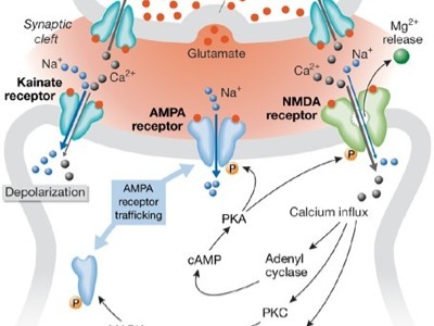
Glutamate is the most abundant neurotransmitter in the brain and operates
through a very complex series of receptors (3 major inotropic receptors- NMDA,
AMPA and kainite receptors, and 8 metabotropic receptors). As stated, the
presence of glutamate outside brain neurons, even in very small concentrations,
is brain cell toxic. Because of this, the brain is equipped with a very
elaborate series of mechanisms to remove glutamate quickly, primarily by
utilizing glutamate uptake proteins (EAAT1-5). Mercury, aluminum, free radicals,
lipid peroxidation products and inflammatory cytokines can easily damage these.
130,131
One of the important ways glutamate regulates neuron function is by allowing
calcium to enter the cell and by the release of calcium within cell storage
depots. When calcium (glutamate operated) channels are opened, the calcium flows
in as a wave of concentrated calcium. These are referred to a calcium waves or
oscillations. They regulate a number of neuron functions, one of which plays a
vital role in brain development.
During brain development, the future neurons are lined up along membranes within
the core of the undeveloped brain. These cells must migrate outwardly to reach
their final destination and they do so by guided chemical signals mainly
released by microglia and astrocytes. These trillions of connections also
develop during a process called synaptogeneis, and use many of the same signals.
Studies have shown that the calcium waves cause developing brain cells to
migrate, which is essential for development of the brain (it forms the
architectonic structures and functional columns of the brain).132 Interestingly,
testosterone also affects embryonic brain cell migration by regulating calcium
waves, and mercury, probably by stimulating glutamate release, does the same
thing.133 Estrogen reduces calcium oscillations and stops the migration. Other
chemical signals in the brain also play a role (reelin).
If calcium oscillations are not properly regulated, that is- there are too many
calcium oscillations, the brain develops abnormally. Testosterone and glutamate
have an additive effect on these calcium waves. In this way, testosterone
enhances the damaging effect of excessive glutamate and mercury.
Studies have shown that higher doses of MSG during brain formation can cause
abnormalities of brain development that closely resemble mercury poisoning and
the toxic effects of high levels of inflammatory cytokines.76 Interestingly,
vaccination has been shown to significantly increase the toxicity of several
other neurotoxins, so much so that they can trigger brain cell destruction or
synaptic loss even when subtoxic concentrations of the toxicants are used.
Testosterone aggravates this toxicity as well.
Studies of autistic children show an elevated level of androgens in most, even
in female autistic children.134 In general, androgens, such as testosterone,
enhance neurological injury and estrogens tend to be protective of the brain.135
The Role of the Leaky Gut Phenomenon and Food Intolerances.
Wakefield and his co-workers demonstrated a connection between the MMR vaccines
and abnormal gut function in a landmark article appearing in the journal Lancet
in 1998.136 In this carefully conducted study they biopsied the lining of the
intestines of autistic children having GI symptoms and demonstrated lymphocytic
infiltration as well as elevated levels of inflammatory antibodies and
cytokines. TNF- release was particularly high from these gut-based immune cells.
The entire GI tract, from the stomach to the colon, was infiltrated by these
immune cells.
Subsequent studies have shown a high incidence of abdominal pain, bloating,
diarrhea and constipation in children with ASD.138,139 A number of other studies
have shown problems with digestive enzymes, defective detoxification, and an
overgrowth of a number of pathogenic bacteria and fungi in the colon and
intestine of ASD children.140,141
Not surprisingly, a few studies have shown significant improvement in behavior
when ASD children are placed on diets devoid of identified food
allergens.142-144 Antibodies to food components, such as casein, gliadin and
gluten have also been described as well as cross-reactions between food antigens
and brain components.145
One disease that closely resembles the case of ASD in terms of brain injury
associated with food allergins is celiac disease, in which there is an immune
sensitivity to the food components gliadin and gluten. Approximately 6% of such
patients will demonstrate neurological damage, most frequently cerebellar
ataxia.146 Other studies have also found seizures, cranial nerve damage,
dementia and impaired frontal lobe function.147-151
Autopsy studies indicate that the most commonly found neurological damage occurs
in the cerebellum, as we see in autism. Other studies have shown an immunologic
cross-reactivity between gluten antibiodies and Purkinje cells in the
cerebellum.144 Like the celiac cases, in autism the most intense microglia
activation and neuronal loss occurred in the cerebellum. In many of the cases of
autistic brains examined, virtually all of the Purkinje cells were lost.54
Studies looking for the incidence of GI symptoms in autistic children indicate
that from 20% to 84% will have complaints. It is interesting to note that in the
studies on celiac-related neurological problems, only 13% complained of GI
symptoms, so ASD children can have gut-related brain effects without obvious GI
symptoms.154
Some feel that the gliadin, casein and gluten can be converted to opioid-like
substances, such as gliadomorphin and casomorphin that can produce a morphine
response in the brain, leading to abnormal behavior.152 These opioids also
suppress immunity and increase excitotoxicity.154 While the opioid effect
exists, I feel it is the recurrent immune stimulation of primed microglia that
is causing most of the damage seen in autism.155
Studies have also found frequent dysbiosis in autistic children, that is, an
overgrowth of pathogenic bacteria and fungi and a loss of beneficial probiotics
organisms.138 It has been demonstrated that Candida organisms can penetrate the
gut wall and enter the blood stream, were they can be distributed to all tissues
and organs, including the brain.156 The same is true for pathogenic bacteria and
bacterial toxins. These brain implanted organisms act as continuous sources of
immune stimulation, which is especially damaging to the brain because of
vaccine-triggered microglia priming and/or activation occurring before the gut
problem presents itself, with repeated vaccination aggravating the injury.
With each subsequent vaccination, the microglia response is enhanced because of
the recurrent immune activation by food antigens and microbiological antigens.
It is interesting to note that trials of antibiotic vancomycin, which is not
absorbed from the gut, objectively improved the cognitive function of a number
of autistic children.157 We also know that with children having celiac disease
even a very small amount of the offending food can have devastating neurological
effects.
Conclusion
I have presented a considerable amount of evidence for a connection between the
present vaccine schedule and the development of autism spectrum disorders, yet
even this paper is only a brief review of what we know. A more in-depth
discussion of the immune/excitotoxic will appear in my paper- Interaction of
activated microglia, excitotoxicity, reactive oxygen and nitrogen species, lipid
peroxidation products and elevated androgens in autism spectrum disorders,
which will appear in an upcoming special autism issue of the journal-Alternative
Therapies in Health and Medicine.
Much of this information is being totally ignored by the medical elite and
especially the media. The Simpsonwood conference proceedings, in which over 50
scientists, vaccine pharmaceutical company representatives and representatives
from the World Health Organization met secretly in Norcross, Georgia, disclosed
that the safety of your children is not their primary interest –their only
interest is selling vaccines to the public. A friend of mine, while speaking to
an audience of scientists and public health officials in Italy, was rudely told
by a public health official that (paraphrased) –We all know that vaccines can
cause neurological damage, but we must keep this from the public because it
might endanger the vaccine program.
It is also important to understand that most practicing pediatricians have never
heard what I have disclosed to you. Most have very little understanding of
immune function and have no idea of the pathological effect on the brain of
giving multiple vaccines on a large scale. These effects are widely discussed in
the neuroscience literature, but few practicing physicians, especially
pediatricians, ever read such articles.
Immunology, like nutrition, gets only scant attention in medical school and even
less in residency training of physicians. Older doctors have no concept of the
newer discoveries in immunology, especially neuroimmunology. The human immune
system is one of the most complex systems in physiology and our studies indicate
an even greater complexity is to be found. Despite a renewed interest in the
immune system’s function in neonates and small children, much remains unknown
concerning the immune effects of exposing infants and small children to such a
large barrage of vaccine early in life. Yet, what we do know is that they react
quite differently than adults and it can have devastating consequences on brain
development and function.
Vaccinating millions of children with the hepatitis B vaccine at birth can only
be described as dangerous idiocy. The vast majority of infants, children and
adolescents are in no danger from this infection- even the medical authorities
agree on that. It is also known that the effectiveness of the vaccine in
children last no more than two years and has little or no effectiveness in the
immune suppressed child. The nefarious plan by these vaccine geniuses is to
force vaccines all babies, since they would have difficulty convincing adults,
that is, the one at any danger, to get the vaccine.
The problem with this “plan” is that the vaccine is ineffective by the time the
child reaches the age of risk. Now that they have discovered this, they are
recommending that all children have a booster vaccine every two years.
The American Academy of Pediatrics and the CDC, the forces behind this vaccine
mania, assure parents that giving all of the required vaccines at once is
perfectly safe. As we have seen, the scientific “evidence” does not support this
policy. To do so exposes the child to a high concentration of immune-stimulating
adjuvants that will intensely activate the brain’s immune system (microglia)
during the brain’s most active growth period, that is, during the first 2 to 6
years of life. The maturation and development of the brain continues to a large
degree throughout adolescence. As we have seen, excessive vaccination can result
in brain inflammation and brain swelling that can be prolonged, even lasting
years, if not decades (as we have seen in the Vargas et al study). This can
result in seizures, high pitched crying, severe lethargy, weakness and
behavioral problems, such as agitation, depression, anger and other autistic
behaviors.
In addition, giving the vaccines all at once exposes the brain to higher levels
of neurotoxic aluminum as proven by radiolabeled aluminum study quoted above. If
a person were to follow recommended vaccine guidelines they would receive over
100 vaccines in a lifetime. Because of the way the vaccines are given, this
would not allow the brain’s microglial cells to shut down, which is essential.
One of the effects of chronic microglial activation, other than brain
inflammation, is an elevation in brain glutamate levels. Studies have shown this
can lead to chronic neurodegeneration and is suspected as a common mechanism
associated with neuropathic viruses, such as the measles and borna
viruses.158-160 In fact, blocking certain of the glutamate receptors can prevent
brain damage by the measles virus, as well as other viruses.158 We also know
that the prognosis of spinal meningitis can be determined by the spinal fluid
glutamate levels, with high levels having the worst prognosis.161 Studies of
autistic children have also shown elevated glutamate levels in their blood and
spinal fluid.
Because excitotoxicity plays such an important role in autism, parents of
autistic children should avoid feeding their children foods containing
excitotoxic additives, such as MSG, hydrolyzed protein, vegetable protein
extracts, soy protein or soy protein isolate, natural flavoring, yeast enzymes,
etc. There are many disguised names for high glutamate food additives. A recent
study has also shown that there is an interaction between certain food dyes and
glutamate and aspartame that enhances neurotoxicity significantly.
They should also avoid immune suppressing oils, such as the omega-6 oils (corn,
soybean, peanut, safflower, sunflower and peanut oils). As stated, people in
this country eat 50-times the amount of this immune suppressing oil than they
need for health.
While omega-3 oils are healthy, the EPA component is significantly immune
suppressing and as a result, high intakes should be avoided. Studies have shown
suppressed lymphocyte function (NK cells) with high intake of EPA.162 It is the
DHA component that has most of the beneficial effects, especially as regards
brain repair and inflammation reduction.163 DHA also inhibits excitotoxicity.
Because the autistic child has intense brain inflammation, a combination of EPA
and DHA is preferable, with a lower content of EPA (no more than 250 mg).
Milk and milk products should be avoided and foods containing gliadin and gluten
should also be avoided. Soy foods are also responsible for a significant number
of food allergies as well as being very high in glutamate, fluoride and
manganese. Fluoride should be avoided, especially in drinking water. Water is
also a significant source of aluminum in the diet (it is added as a clarifying
agent) and in fluoridated water the fluoride complexes with aluminum to form the
highly neurotoxic fluoroaluminum compound. The greatest dietary source of
aluminum is biscuits, pancakes, black tea and baked goods made with aluminum-containing
baking powder.
Low magnesium intake, which is common in the United States, is associated with
higher degrees of inflammation in the body and lower glutathione levels. It also
enhances excitotoxicity, since magnesium is a natural modulator of the NMDA
glutamate receptor. Low intakes of magnesium greatly enhance glutamate receptor
sensitivity, worsening excitotoxicity. Low magnesium also lowers brain
glutathione levels, which increases brain sensitivity to mercury toxicity.
Increasing magnesium levels, reduces inflammation, raises glutathione levels and
reduces excitotoxic sensitivity.
A number of flavonoids are neuroprotective, especially against inflammation and
excitotoxicity. These include curcumin, quercetin, ellagic acid, natural vitamin
E (mixed trocopherol), epigallocatechin gallate (from white tea), theanine, DHEA
and hesperidin. All are available as supplements and most have a high safety
profile.
The live virus vaccines, such as chickenpox, measles, mumps and rubella, pose a
special danger in the immunosuppressed child, because some of these viruses can
take up permanent residence in the body, including the brain. In one study,
which examined the tissues of elderly dying of non-infectious causes, found live
measles virus in 45% of the bodies examined and 20% of their brains.164,165
These measles viruses were highly mutated, meaning they could result in a number
of diseases not normally suspected with measles infection.
I have omitted discussions about vaccine contamination, which is a major
problem. Several studies found a high incidence of microorganism contamination
in vaccines made by a number of major pharmaceutical companies, with figures as
high a 60% of the vaccines being contaminated.94-99 Bacterial and viral
fragments have also been found in a number of vaccines. While vaccine promoters
were quick to assure us that these viral fragments should cause no problem,
research says otherwise. In fact, a non-viable viral fragment implanted in
microglia and astrocytes in the brain causes the devastating dementia associated
with the HIV virus.167,168 The virus does not infect the brain neurons
themselves. The mechanism proposed is an immunological/excitotoxic-induced
toxicity, just as we see with repeated vaccination. The same mechanism is seen
with a number of viruses, including measles viruses, borna virus and the herpes
virus.168-172
When brain glial cells or neurons are chronically infected with these viruses
(called a persistent viral infection) the smoldering immune/excitotoxic reaction
slowly destroys the brain cell connections because the immune system is
attempting to destroy the infectious microorganism. Since it can never kill the
organism, the destruction (and intense microglial activation) continues for
decades, as we saw in the autistic brain.54 The same can occur with viral
fragments, the Lyme disease organism, aluminum and mercury that has accumulated
in the brain from either contaminated vaccines or from vaccine additives. And
because excessive vaccination, especially with immune-suppressive viruses, can
depress proper immune function, the child is at a greater risk of developing
such a persistent viral infection. Likewise, they are at a greater risk of
developing deadly invasive bacterial infections, such as H. Influenza
meningitis, pneumococcal and meningiococcal meningitis. When it occurs, the
vaccine promoters scream that we need more vaccines to protect the children,
never admitting that it was the vaccine program itself that destroyed the lives
of these children.
While a number of people and even physicians, think they desire a universal
health care system (a euphemism for socialized medicine), here is something to
consider. The government will use access to health care as a way to mandate
vaccinations for all Americans. Those who refuse any of the mandated vaccines
will be denied access to health care, meaning you will not be able to see a
doctor or enter a hospital or clinic.
All federal programs will have completion of vaccine mandates as a requirement.
This could be linked to social security, food stamps, housing subsidy programs
and other such federal programs. Remember, they use such tactics now for access
to schools and daycare centers. One may even have to prove that they have had
all their required vaccinations before they can use public transportation, such
as busses, trains and airplanes.
Another thing to consider is that the communist Chinese are gradually taking
over vaccine manufacturing. In fact, communist China is now the largest vaccine
manufacturer in the world . They have over 400 biopharmaceutical companies busy
making vaccines and poor quality drugs for the world. The FDA admits that it
inspects only 1.8% of the 714 drug firms in China and that, according to a GAO
study, that FDA inspections may be 13 years apart (it is spaced 2 years apart in
the United States).
Even more frightening is that the inspectors must depend on Chinese translators
and US companies purchasing these vaccines and pharmaceuticals must, by
agreement, have a Chinese communist official serve as its legal representative.
According to the Phyllis Schafly Report, one CEO was quoted as saying “ every
piece of information you get (from the Chinese) is suspect.”
With thousands of people dying and getting sick, not only in China, but in
hundreds of nations receiving their tainted pharmaceutical products, this means
future vaccines will be an even greater danger. The risk of millions of
Americans and others living in the West receiving contaminated vaccines is
extremely high. It could even be done on purpose, since the Chinese communist
have declared their intention to defeat the United States. Infecting over a
hundred million Americans with contaminated vaccines would be an easy way to
defeat us. The irony would be that our public officials would have aided them in
our destruction.
Parents must appreciate that those in positions of authority are lying to them.
Most pediatricians think they are doing what is right, because they too are
victims of years of propaganda by elite members in the CDC and American Academy
of Pediatrics. Most truly believe what they are telling parents. They should
wake up and joint the fight to bring some sense to this insane policy.
References
1.Money J et al. Autism and autoimmune disease: A family study. J Autism Child
Schizophr 1971; 1: 146-160.
2.Comi A. et al. Familial clustering of autoimmune disorders and evaluation of
medical risk factors in autism. J Child Neurology 1999; 14: 388-394.
3.Sweetwen TL et al. Increased prevalence of familial autoimmunity in probands
with pervasive developmental disorders. Pediatrics 2003: 112: 420.
4.Creen LA et al. Maternal autoimmune disease, asthma and allergies, and
childhood autism spectrum disorders: a case-control study. Arch Pediatr
2005;159: 151-157.
5.Dalton P et al. Maternal antibodies associated with autism and language
disorders. Ann Neurol 2003;53: 533-537.
6.Singh VK, Rivas WH. Prevalence of serum antibodies to caudate nucleus in
autistic children. Neuroscience Lett 2004; 355: 53-56.
7.Singh VK et al. Antibodies to myelin basic protein in children with autistic
behavior. Brain Behavior Immunol 1993; 7: 97-103.
8.Singer HS et al. Antibrain antibodies in children with autism and their
unaffected siblings. J Neuroimmunol 2006; 178: 149-155.
9.Singh VK et al. Circulating autoantibodies to neural and glial filament
proteins in autism. Pediatr Neurol 1997; 17: 88-90.
10.el-Fawal HA e al. Exposure to methylmercury results in serum autoantibodies
to neurotypic and gliaotypic proteins. Neurotoxicology 1996; 17: 531-539.
11.Havarinasab S et al. Immunosuppressive and autoimmune effects of thimerosal
in mice. Toxicol Appl Pharmacol 2005; 204; 109-121.
12.Hornig M, Chian D, Lipkin WJ. Neurotoxic effect of postnatal thimerosal are
mouse strain dependent. Mol Psychiatry 2004; 9: 833-845.
13.Tishler M, Shoenfeld Y. Vaccination may be associated with autoimmune
disease. Isr Med Assoc J 2004; 6: 430-432.
14.Shoenfeld T, Aron-Maor A. Vaccination and autoimmunity-‘vaccinosis’ a
dangerous liaison? J Autoimmunity 2000; 14: 1-10.
15.Vojdam A et al. Antibodies to neuron-specific antigens in children with
autism: possible cross-reaction with encephalitogenic proteins from milk,
Chlamydia pneumoniae and Streptococcus group A. J Neuroimmunol 2002; 129:
168-177.
16.Lucarelli S et al. Food allergy and infantile autism. Panminerva Med 1995;
37: 137-141.
17.O’Banion D et al. Disruptive behavior: a dietary approach. J Autism Child
Schizophr 1978; 8: 325-337.
18.Vojdani A et al. Immune response to dietary proteins, gliadin and cerebellar
peptides in children with autism. Nutr Neuroscience 2004; 7: 151-161.
19.McGeer PL and McGeer EG. Autotoxicity and Alzheimer Disease. 2000; 57;
289-290.
20.Malek-Ahmadi P. Cytokines and etiopathogenesis of pervasive developmental
disorders. Med Hypothesis 2001; 56: 321-324.
21.Weizman A et al. Abnormal responses to brain tissue antigen in the syndrome
of autism. Am J Psychiatry 1982; 139; 1462-1465.
22.Lee SC et al. Cytokine production by human fetal microglia and astrocytes.
Differential induction by liposaccharide and IL-1beta. J Immunol 1993; 150:
2659-2667.
23.Bauer S et al. The neuropoetic cytokine family in development, plasticity,
disease and injury. Nature Reviews/Neuroscience 2007; 8: 221-232.
24.Boulanger LM, Shatz CJ. Immune signaling in neural development, synaptic
plasticity and disease. Nature Reviews/Neuroscience 2004; 5: 521-531.
25.Agrawal A et al. Thimerosal induces TH2 responses via influencing cytokine
secretion by human dendritic cells. J Leukocyte Biol 2007; 81: 1-9.
26.Kidd P. Th1/Th2 balance: The hypothesis, its limitations, and implication in
health and disease. Altern Medicine Rev 2003; 8: 223-246.
27.Martin OC et al. Hepatitis B immunization induces higher antibody and memory
Th2 responses in new-borns than adults. Vaccine 2004; 22: 511-519.
28.Cohly HH, Panja A. Immunologic findings in autism. In Rev Neurobiol 2005; 71:
317-341.
29.Singh VK. Plasma increase of interleukin-12 and interferon-gamma.
Pathological significance in autism. J Neuroimmunol 1996; 66: 143-145.
30.Jyonouchi H et al. Proinflammatory and regulatory cytokine production
associated with innate and adaptive immune responses in children with autism
spectrum disorders and developmental regression. J Neuroimmunol 2001; 120:
170-179.
31.Pandey RS et al. Autoimmune model of schizophrenia with special reference to
antibrain antibodies. Biol Psychiatry 1981;16: 1123-1136.
32.Zhang XY et al Elevated interleukin-2, interleukin-6 and interleukin-8 serum
levels in neuroleptic-free schizophrenia: association with psychopathology.
Schizophr Res 2002; 57: 247-258.
33.Turner W et al. Measles associated encephalopathy in children with renal
transplants. Am J Transplant 2006; 6: 1459-1465.
34.Larner AJ, Farmer SF. Myelopathy following influenza vaccination in
inflammatory disorder treated with chronic immunosuppression. Eu J Neurol 2000;
7: 731-733.
35.Kerdile YM et al. Immunosuppression by measles virus: role of viral proteins.
Rev Med Virol 2006; 16: 49-63.
36.Abernathy RS, Spink WW. Increased susceptibility of mice to bacterial
endotoxins induced by pertussis vaccine. Fed Proc 1956; 15: 580.
37.Auwaerter PD et al. Changes within T-cell receptor V beta subsets in infants
following measles vaccinations. Clin Immunol Immunopathol 1996; 79: 163-167.
38.Hussey GD et al. The effect of Edmonston-Zagreb and Schwartz measles vaccines
on immune responses in infants. J Infect Dis 1996; 173: 1320-1326.
39.Hirsch RL et al. Measles virus vaccination of measles seropositive
individuals suppresses lymphocyte proliferation and chemotactic factor
production. Clin Immunol Immunopath 1981; 21: 341-350.
40.Daum RS et al. Decline in serum antibody to the capsule of Haemophilus
influenza type b in the immediate postimmunization period. J Pediatrics
1989;1114: 742-747.
41.Pukhalsky AL et al. Cytokine profile after rubella vaccine inoculation:
evidence of the immunosuppressive effect of vaccination. Mediators Inflammation
2003; 12: 203-207.
42.Miller NZ. Vaccine Safety Manuel: For Concerned Families and Health
Practioners. New Atlantean Press, NM, 2008.
43.Pichichero ME et al. Pathogen shifts and changing cure rates for otitis media
and tonsillopharyngitis. Clin Pediatr 2006; 45: 493-502.
44.Moore MR et al. Impact of conjugate vaccine on community wide coverage of
nonsusceptible Streptococcus in Alaska. J Inf Dis 2004; 190: 2031-2038.
45.Pichichero ME, Cary JR. Emergence of a multiresistant serotype 19A
pneumococcal strain not included in the 7-valent conjugate vaccine as an
otopathogen in children. JAMA 2007; 298: 1772-1778.
46.Strunecka A., Patocka J, Blaylock RL et al. Fluoride interactions: From
molecules to disease. Current Signal Transduction Therapy 2007; 2
47.Block ML, Zecca L, Hong J-S. Microglia-mediated neurotoxicity: uncovering the
molecular mechanisms. Nature Reviews/Neuroscience 2007; 8: 57-69.
48.Mandu P, Brown GC, Activation of microglial NADPH oxidase is synergistic with
glial NOS expression in inducing neuronal death: a duel-key mechanism of
inflammatory neurodegeneration. 2005; 2: 20.
49.Cagnin A et al. In vivo visualization of activated glia by [11C] (R)-
PK11195-PET following herpes encephalitis reveals projected neuronal damage
beyond the primary focal lesion. Brain 2001; 124: 2014-2027.
50.Lemstra AW et al. Microglia activation in sepsis: a case-control study. J
Neuroinflamm 207; 4: 4
51.Buttini M, Lumonta S, Boddeke HW. Peripheral administration of
lipopolysaccharide induces activation of microglial cell in rat brain. Neurochem
Int 1996; 29: 25-35.
52.Cunningham C et al. Central and systemic endotoxin challenges exacerbate the
local inflammatory responses and increased neuronal death during chronic
neurodegeneration. J Neurosci 2005; 25: 9275-9284.
53.Godbout JP et al. Exaggerated neuroinflammatory and sickness behavior in aged
mice following activation of the peripheral innate immune system. FASEB J
2005;19: 1329-1331.
54.Vargas DL et al. Neuroglial activation and neuroinflammation in the brain of
patients with autism. Ann Neurol 2005; 57: 67-81.
55.Blaylock RL. Central role of excitotoxicity in autism. JANA 2003;6: 7-19.
56.Lewine JD et al. Magnetoencephalographic patterns of epileptiform activity in
children with regressive autism spectrum disorders. Pediatrics 1999; 104:
405-415.
57.Auvin S et al. Inflammation exacerbates seizure-induced injury in the
immature brain. Epilepsia 2007; 48: 27-34.
58.Rizzi M et al. Glia activation and cytokines increased in rat hippocampus by
kainic acid-induced status epilepticus during postnatal development. Neurobiol
Dis 2003; 4: 94-103.
59.Eastman CL et al. Increased brain quinolinic acid production in mice infected
with hamster neurotropic measles virus. Exp Neurol 1994; 125: 119-124.
60.Heyes MP et al. Human microglia convert L-tryptophan into neurotoxin
quinolinic acid. Biochem J 1996; 320: 595-597.
61.Ida T et al. Cytokine-induced enhancement of calcium-dependent glutamate
release from astrocytes mediated by nitric oxide. Neurosci Lett 2008; 432:
232-236.
62.Ye GL et al. AMPA and NMDA receptor-mediated currents in developing dentate
granule cells. Brain Res Dev Brain Res 2005; 155: 26-32.
63.Menkes JH, Kinsbourne M. Workshop on neurologic complications of pertussis
and pertussis vaccinations. Neuropediatrics 1990; 21: 171-176.
64.Kiviravanta T, Airaksinen EM. Low sodium levels in serum are associated with
febrile seizures. Acta Paediatr 1995; 84: 1372-1374.
65.Bar-Peled O et al. Distribution of glutamate transporter subtypes during
human brain development. J Neurochem 1997; 69: 2571-2580.
66.Arauz-Contreas J, Feria-Velasco A. Monosodium-L-glutamate-induced convulsions
1. Differences in seizure pattern and duration of effect as a function of age in
rats. Gen Pharmacol 1984; 15: 391-395.
67.Neil Z. Miller. Vaccines: Are they Really Safe and Effective? A Parent’s
Guide to Childhood Shots. New Atlantean Press, NM 1999.
68.Toga Aw et al. Mapping brain maturation. Trend Neurosci 2006; 29: 148-159.
69.Gogtay N et al. Dynamic mapping of human cortical development during
childhood and adolescence. Proc Natl Acad Sci USA 2006; 101: 8174-8179.
70.Jerigan TL, Tallal P. Late childhood changes in brain morphology observable
with MRI. Dev Med Child Neurol 1990; 32: 379-385.
71.Maslinska D et al. Morphological forms and localizations of microglial cells
in the developing human cerebellum. Folia Neuropathol 1998; 36: 145-151.
72.Monier A et al. Entry and distribution of microglial cells in human embryonic
and fetal cerebral cortex. J Neuropathol Exp Neurol 2007; 66: 372-382.
73.Schwab JM et al. IL-6 is differentially expressed in the developing human
fetal brain by microglial cells in zones of neuropoesis. In J Dev Neurosci 2001;
114: 232-241.
74.Schlett K. Glutamate as a modulator of embryonic and adult neurogenesis. Curr
Top Med Chem 2006; 6: 949-960.
75.Kumuro H, Rakic P. Modulation of neuronal migration by NMDA receptors.
Science 1993; 260: 95-97.
76.Marret S et al. Arrest of neuronal migration by excitatory amino acids in
hamster developing brain. Proc Natl Acad Sci USA 1996; 93: 15463-15468.
77.Aarum J et al. Migration and differentiation of neural precursor cells can be
directed by microglia. Proc Natl Acad Sci USA 2003;100: 15983-15988.
78.Ekdahl CT et al. Inflammation is detrimental for neurogenesis in adult
brains. Proc Natl Acad Sci USA 2003; 100: 13632-13635.
79.Chao CC et al. Tumor necrosis factor-alpha potentates glutamate neurotoxicity
in human fetal cell cultures. Dev Neurosci 1994; 16: 172-179.
80.Kemper TL et al. Neuropathology of infantile autism. J Neuropathology Exp
Neurol 1998; 57: 645-652,
81.Bauman MI, Kemper TL. The neuropathology of autism spectrum disorders: What
have we learned? Novartis Foundation Symp 2003; 251: 112-122.
82.Bauman M, Kemper TL. Developmental cerebellar abnormalities: a consistent
finding in early infantile autism. Neurology 1986; 36 (Suppl 1): 190.
83.Courchesne E. Brainstem cerebellar and limbic neuroanatomical abnormalities
in autism. Curr Opin Neurobiol 1997; 7: 269-278.
84.Buller KM, Day TA. Systemic administration of interleukin 1beta activates
select populations of central amygdala afferents. J Comp Neurol 202; 452:
288-296.
85.Taylor DL et al. Stimulation of microglial metabotropic glutamate receptor
mGlu2 triggers tumor necrosis factor -induced neurotoxicity in concert with
microglial-derived Fas ligand. J Neurosci 2005; 25: 2952-2964.
86.Rothwell NJ. Cytokines-Killers in the brain? J Physiol 1999; 514.1: 3-17.
87.Samland H et al. Profound increase in sensitivity to glutamatergic –but not
to cholinergic agonist-induced seizures in transgenic mice with astrocytes
production of IL-6. J Neurosci Res 2003; 73: 176-187.
88.Bernardino L et al. Modulator effects of interleukin-1ß and Tumor necrosis
factor- on AMPA-induced excitotoxicity in mouse organotypic hippocampal slice
cultures. J Neurosci 2005; 25: 6734-6744.
89.Allan SM et al. Interleukin-1 and neuronal injury. Nature Reviews/Immunol
2005; 5: 629-640.
90.Burka SL et al. Maternal cytokine levels during pregnancy and adult
psychosis. Brain Behav Immunol 2001; 15: 411-420.
91.Brown AS et al. Elevated maternal interleukin-8 levels and risk of
schizophrenia in adult offspring. Am J Psychiatry 2004; 161: 889-895.
92.Ganguli R et al. Autoimmunity in schizophrenia: a review of recent findings.
Ann Med 1993; 25: 489-496.
93.Schori H et al. Severe immunodeficiency has opposite effects in neuronal
survival in glutamate-susceptible and resistant mice: adverse effect of B-cells.
J Immunol 2002; 169: 2861-2865.
94.Cutrone R et al. Some oral polio vaccines were contaminated with infectious
SV-40 after 1961. Can Res 2005; 65: 10273-10279.
95.Harasawa R, Tomiyama T. Evidence of pestivirus RNA in human virus vaccines. J
Clin Microbiol 1994; 32: 1604-1605.
96.Geier M et al. Endotoxins in commercial vaccines. Appl Environ Microbiol
1978; 36: 445-449.
97.Giangaspero M et al. Genotypes of pestivirus RNA detected in live virus
vaccines for human use. J vet Med Sci 2001; 63: 723-733.
98.Potts BJ et al. Possible role of pestivirus in microcephaly. Lancet 1987;1:
972-973.
99.Johnson JA, Heneine W. Characteristics of endogenous avian leukosis virus in
chicken embryonic fibroblast substrates used in production of measles and mumps
vaccine. J Virol 2001; 75: 3605-3612.
100.Gherardi RK et al. Macrophagic myofasciitis lesion assess long-term
persistence of vaccine-derived aluminum hydroxide in muscle. Brain 2001; 124:
1821-1831.
101.Authier F-J et al. Central nervous system disease in patients with
macrophagic myofasciitis. Brain 2001; 124: 974-983.
102.Bonnefont-Rousselot D et al. Blood oxidative status in patients with
macrophagic myofasciitis. Biomed Pharmacol 2004; 58: 516-519.
103.Good PF et al. Selective accumulation of aluminum and iron in the
neurofibrilary tangles of Alzheimer’s disease: a laser microprobe (LAMMA) study.
Ann Neurol 1992; 31: 286-292.
104.Esparza JL et al. Aluminum-induced pro-oxidant effect in rats: protective
role of exogenous melatonin. J Pineal Res 2003; 35: 32-39.
105.Yokel RA et al. The distribution of aluminum into and out of the brain. J
Inorg Biochem 1999; 76: 127-132.
106.Campbell A et al. Chronic exposure to aluminum in drinking water increases
inflammatory parameters selectively in the brain. J Neuroscience Res 2004; 75:
565-572.
107.Bishop NJ et al. Aluminum neurotoxicity in preterm infants receiving
intravenous feeding solutions. N Engl J Med 1997; 336: 1557-1561.
108.Campbell A. Inflammation, neurodegenerative disease, and environmental
exposures. Ann NY Acad Sci 2004; 1035: 117-132.
109.Shirabe T et al. Autopsy case of aluminum encephalopathy. Neuropathology
2002; 22: 206-210.
110.Armstrong RA et al. Hypothesis: Is Alzheimer’s disease a metal-induced
immune disorder. Neurodegeneration 1995; 4: 107-111.
111.Flarend RE et al. In vivo absorption of aluminum-containing vaccine
adjuvants using 26Al. Vaccine 1997; 15: 1314-1318.
112.Platt B et al. Aluminum toxicity in the rat brain: histochemical and
immunocytochemical evidence. Brain Res Bull 2001; 55: 257-267.
113.Brookes N. Specificity and reliability of the inhibition by HgCl2 of
glutamate transport in astrocytes cultures. J Neurochem 1988; 50: 1117-1122.
114.Vahter ME et al. Demethylation of methylmercury in different brain sites of
Macaca fascicularis monkeys during long-term subclinical methylmercury exposure.
Toxicol Appl Pharmacol 1995; 134: 273-284.
115.Charleston JS et al. Changes in the number of astrocytes and microglia in
the thalamus of the monkey Macaca fascicularis following long-term subclinical
methylmercury exposure. Neurotoxicology 1996; 17: 127-138.
116.Charleston JS et al. Increase in the number of reactive glia in the visual
cortex of Macaca fascicularis following subclinical long-term methylmercury
exposure. Toxicol Appl Pharmacol 1994; 129: 196-206.
117.Burbacher TM et al. Comparison of blood and brain mercury levels in infant
monkeys exposed to methylmercury or vaccines containing thimerosal. Environ
Health Perspect 2005; 113: 1015-1021.
118.Mutkus L et al. Methylmercury alters the in vitro uptake of glutamate and
GLAST and GLT-1 transfected mutant CHO-K1 cells. Biol Trace Elem Res 2005; 107:
231-245.
119.Aschner M et al. Methymercury alters glutamate transport in astrocytes.
Neurochem Int 2000; 37: 199-206.
120.Kim P, Choi BH. Selective inhibitors of glutamate uptake by mercury in
cultured mouse astrocytes. Yonsi Med J 1995; 36: 299-305.
121.Kugler P, Schleyer V. Developmental expression of glutamate transporters and
glutamate dehydrogenase in astrocytes of the postnatal rat hippocampus.
Hippocampus 2004; 14: 975-985.
122.Yel L et al. Thimerosal induces neuronal cell apoptosis by causing
cytochrome C and apoptosis-inducing factor release from mitochondria. In J Mol
Med 2005; 16: 971-977.
123.Humphrey ML et al. Mitochondria mediated thimerosal-induced apoptosis in a
human neuroblastoma cell line (SK-N-SH). Neurotoxicology 2005; 26: 407-416.
124.Henneberry RC. The role of neuronal energy in neurotoxicity of excitatory
amino acids. Neurobiol Aging 1989; 10: 611-613.
125.Zeevalk GD et al. Excitotoxicity and oxidative stress during inhibition of
energy metabolism. Dev Neurosci 1998; 20: 444-445.
126.Haley BE. The relationship of the toxic effects of mercury to exacerbation
of the medical condition classified as Alzheimer’s disease. Medical Veritas
2007; 4: 1510-1524.
127.Sun YM et al. Sex-specific impairment in sexual and ingestive behaviors of
monosodium glutamate-treated rats. Physiol Behavior 1991;50: 873-880.
128.Yang S-H et al. Testosterone increases neurotoxicity of glutamate in vitro
and ischemia-reperfusion injury in an animal model. J Appl Physiol 2002; 92:
195-201.
129.Estrada M et al. Elevated testosterone induces apoptosis in neuronal cells.
J Biol Chem 2006; 281: 25492-25501.
130.Aschner M et al. Involvement of glutamate and reactive oxygen species in
methyl mercury neurotoxicity. Braz J Med Biol Res 2007; 40: 285-291.
131.Allen JM et al. The consequences of methylmercury exposure on interactive
function between astrocytes and neurons. Neurotoxicology 2002; 23: 755-759.
132.Lautermilch NJ, Spitzer NC. Regulation of calcineurin by growth cone calcium
waves controls neurite extension. J Neurosci 2000; 20: 315-325.
133.Estrada M et al. Ca2+ oscillations induced by testosterone enhance neurite
outgrowth. J Cell Sci 2005; 119; 733-743.
134.Geier DA, Geier MR. A clinical trial of combined anti-estrogen and
anti-heavy metal therapy in autistic disorder. Neuroendocrinol Lett 2006; 27:
833-838.
135.Baker AE et al. Estrogen modulates microglial inflammatory mediator
production via interactions with estrogen receptor ß. Endocrinology 2004; 145:
5021-5032.
136.Wakefield AJ et al. Ileal-lymphoid nodular hyperplasia, non-specific
colitis, and pervasive developmental disorders in children. Lancet 1998; 351:
637-641.
137.Ashwood P, Wakefiled AJ. Immune activation of peripheral blood and mucosal
CD3+ lymphocyte cytokine profiles in children with autism and gastrointestinal
systems. J Neuroimunol 2006; 173: 126-134.
138.Horvath K et al. Gastrointestinal abnormalities in children with autistic
disorder. J Pediatr 1999; 135: 559-563.
139.Afzal N et al. Constipation with acquired megacolon in children with autism.
Pediatrics 2003; 112: 939-942.
140.Feingold SM et al. Gastrointestinal microflora studies in late-onset autism.
Clin Infect Dis 2002; 35: S6-S16.
141.Vojdani A et al. Antibodies to neuron-specific antigens in children with
autism: possible cross-reaction with encephalitogenic proteins from milk,
Chlamydia pneumonia and Streptococcus group A. J Neuroimmunol 2002; 129:
168-177.
142.Lucarelli S et al. Food allergy and infantile autism. Panminerva Med 1995;
37: 137-141.
143.Knivsberg AM et al. A randomized, controlled study of dietary intervention
in autistic syndrome. Nutri Neurosci 2002; 5: 251-261.
144.Vojdani A et al. Immune response to dietary proteins, gliadin and cerebellar
peptides with autism. Nutr Neurosci 2004; 7: 151-161.
145.Whitely P et al. A gluten-free diet as an intervention for autism and
associated disorders: preliminary findings. Autism 1999; m3: 45-65.
146.Bushara KO. Neurologic presentation of celiac disease. Gastroenterology
2005; 128: S92-S97.
147.Kinney HC et al. Degeneration of the central nervous system associated with
celiac disease. J Neurol Sci 1982; 53: 9-22.
148.DeSantis A et al. Schizophrenia symptoms and SPECT abnormalities in a coelic
patient: regression after gluten-free diet. J Intern Med 1997; 242: 421-423.
149.Beyenberg S et al. Chronic progressive leukoencephalopathy in adult celiac
disease. Neurology 1998; 50: 820-822.
150.Burk K et al. Sporadic cerebellar ataxia associated with gluten sensitivity.
Brain 2001; 124: 1013-1019.
151.Hu WT et al. Cognitive impairment and celiac disease. Arch Neurol 2006;63:
1440-1446.
152.Wakefield AJ et al. Review article: The concept of entero-colonic
encephalopathy, autism and opioid receptor ligands. Aliment Pharmacol Ther 2002;
16: 663-674.
153.Peterson PK et al. The opioid-cytokine connection. J Neuroimmunology 1998;
83: 63-69.
154.Zhu L et al. Enhancing effect of beta-endorphins on glutamate toxicity.
Zhongguo Yao Li Xue Bao 1998; 19: 108-111.
155.Blaylock RL. Interaction of cytokines, excitotoxins, and reactive nitrogen
and oxygen species in autism spectrum disorders. JANA 2003; 6: 21-35.
156.Rao S, Ali U. Systemic fungal infections in neonates. J Postgrad Med 2005;
51 (suppl 1): S27-S29.
157.Sandler RH et al. Short term benefit from oral vancomycin treatment of
regressive-onset autism. J Child Neurol 2000; 15: 429-435.
158.Anderson T et al. NMDA-receptor antagonist prevents measles virus-induced
neurodegeneration. Eur J Neurosci 1991; 3: 66-71.
159.Eastman CL et al. Increased brain quinolinic acid production in mice
infected with a hamster neurotropic measles virus. Exp Neurol 1994;125: 119-124.
160.Raslet A et al. Borrelia burgdorferi induces inflammatory mediator
production by murine microglia. J Neuroimmunol 2002; 130: 22-31.
161.Ma W eat al. Elevated cerebrospinal fluid levels of glutamate in children
with bacterial meningitis as a predictor of the development of seizures or other
adverse outcomes. Pediatr Crit care Med 2003; 4: 170-175.
162.Zhao Y et al. Eicopentaenoic acid prevents LPS-induced TNF- expression by
preventing NFkB activation. J Amer Coll Nutr 2004; 23: 71-78.
163.Weldon SM et al. Docosahexaenoic acid induces an anti-inflammatory profile
in liposaccharide-stimulated THP-1 macrophage mice more effectively than
eicosapentaenoid acid. J Nutr Biochem 2007; 18: 250-258.
164.Katayama Y et al. Detection of measles virus nucleoprotein mRNA in autopsied
brain tissue. J Gen Virol 1995; 76: 3201-3204.
165.Katayama Y et al. Detection of measles virus mRNA from autopsied human
tissues. J Clin Microbiol 1998; 36: 299-301.
166.Hult B et al. Neurobiology of HIV. Int Rev Psychology 2008; 20: 3-13.
167.Gonzales-Sarano F, Martin-Garcia J. the neuropathogenesis of AIDS. Nat Rev
Immunol 2005; 5: 69-81.
168.Rubin SA et al. Viral teratogenesis: brain developmental damage associated
with maturation state at time of infection. Brain Dev Rev 1999; 112: 237-244.
169.Lellouch-Tubiana A et al. Immunocytochemical characterization of long-term
persistent immune activation in human brain after herpes simplex encephalitis.
Neuropathology Appl Neurobiol 2000; 26: 285-294.
170.Ovanesov MV et al. Activation of microglia by Borna disease virus infection:
In vitro study. J Virol 2006; 80: 12141-12148.
171.Volmer R et al. Borna disease virus infection impairs synaptic plasticity. J
Virol 2007; 81: 8833-8837.
172.De la Torre JC. Borna virus and the brain. J Infect Dis 2002; 186: (suppl2)
: S241-S247.